- Author Rachel Wainwright wainwright@abchealthonline.com.
- Public 2023-12-15 07:39.
- Last modified 2025-11-02 20:14.
Eyes
A person sees through the eyes, from where the image is transmitted through the chiasm, the optic nerve, the optic tracts to special areas of the occipital lobes of the cerebral cortex. Our vision is three-dimensional due to the presence of two eyes. The right side of the retina transmits the "right side" of the image to the right side of the brain. The same happens with the left side of the retina. And the brain connects these two parts together, getting a whole image.

If the joint movement of the left and right eyes is disturbed, binocular vision will be disturbed, since each eye sees only “its own” image.
Eye functions
- Optical function that projects an image;
- Life support function;
- Perception and "encoding" of the received information for the brain.
Structure
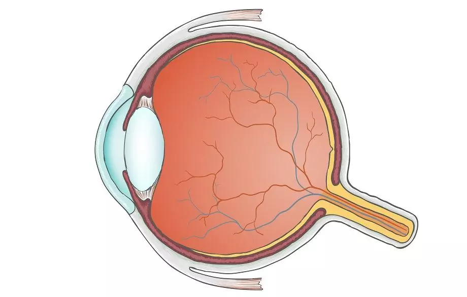
The eye is a complex optical device, the main task of which is to transmit an image to the optic nerve. Currently, the structure of the eye has been studied so well that it allows performing such delicate procedures as laser vision correction.
The eye consists of the following parts:
- The cornea is the clear membrane in the front of the eye. The cornea has no blood vessels, it has a high refractive power and borders on the sclera.
- The anterior chamber is the space between the iris and the cornea filled with intraocular fluid.
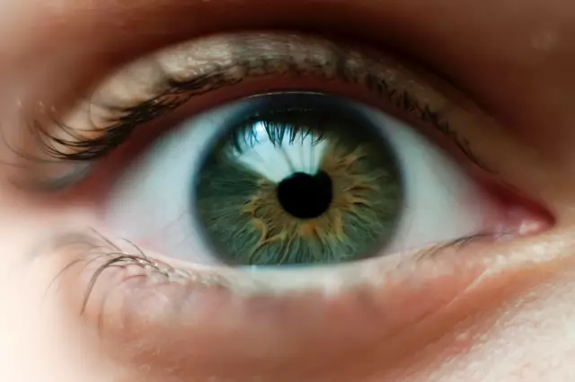
- An iris that is shaped like a circle with a hole. The iris consists of muscles that, when relaxed or contracted, change the size of the pupil. The color of the eyes depends on the iris (if there are few pigment cells on it, it is blue, and if there are many - brown). The iris can be compared to the aperture in a camera that regulates the light flow.
- The pupil is a hole in the iris. The light level affects the size of the pupil. The more light, the smaller the pupil size.
- The lens is a transparent and elastic lens that can easily change its shape, almost instantly "focusing" on something. That is why a person with good vision can see both from close and from a distance. The lens is located in a special capsule, which is held by an eyelash girdle.
- The vitreous humor is a transparent, gel-like substance found in the back of the eye. Its main function is to maintain the shape of the eyeball and participate in intraocular metabolism.
- The retina, which is composed of nerve cells and light-sensitive photoreceptors. Retinal receptors can be divided into two types: rods and cones. They produce the enzyme rhodopsin and convert the light energy into electrical energy of the nervous tissue, that is, a chemical reaction occurs. The sticks have a high light sensitivity and allow a person to see well even in low light. Also, the rods are responsible for peripheral vision. Cones, on the other hand, work well only when there is a lot of light. Thanks to the cones, we can see fine details and distinguish colors.
- The sclera is the outer opaque shell of the eyeball, which in front of it passes into the transparent cornea. Attached to the sclera are six muscles that allow the eye to move. It also contains many vessels and nerve endings.
- Choroid, which is located in the posterior part of the sclera. The membrane is closely connected with the retina and is responsible for the blood supply to the intraocular structures. There are no nerve endings in the choroid, therefore, with diseases of the membrane, a person does not feel pain.
- The optic nerve, through which images are transmitted from nerve endings to the brain.
Eye diseases and treatments
All eye diseases can be divided into infectious and non-infectious.
Infectious diseases arise under the influence of various microorganisms: bacteria, viruses, fungi. It is important for these diseases to undergo a vision test and start adequate eye treatment, otherwise the disease can lead to damage to the cornea and even loss of vision. Infectious diseases include conjunctivitis, blepharitis, and barley.
Non-infectious diseases occur as a result of overexertion of vision, hereditary predisposition, age-related changes. The most common non-communicable diseases include dry eye syndrome, cataracts, and glaucoma.
If non-infectious diseases are detected in the early stages, then eye treatment has favorable prognosis, so it is important to have an eye examination by an ophthalmologist every six months.
Bags and circles under the eyes
In addition to eye diseases, people often have circles and bags under the eyes. Where do they come from?
The fact is that the skin of the eyelids is very delicate and thin. Due to the lack of oxygen or insufficient blood supply, the blood in the capillaries stagnates and begins to shine through. This is how dark circles appear under the eyes.
There are many reasons for their appearance. Circles can be a hereditary factor, a feature of the structure of the subcutaneous fatty tissue, or indicate diseases of the internal organs and the endocrine system. The most common causes of under-eye circles are stress, constant lack of sleep and chronic fatigue.
Puffiness and bags under the eyes often result from drinking a lot of alcohol, tea or coffee before bed. As a result, stagnation of blood and lymph vessels occurs, and as a result - bags over the eyes in the morning.
In addition, bags under the eyes can indicate poor kidney function, an allergic reaction, and inflammation of the sinuses.
Who needs an eye test?
- People with good vision for prevention purposes.
- People who have lost vision. In this case, a comprehensive examination is necessary, as a result of which the doctor may advise to wear glasses, lenses or to carry out a vision correction.
- Contact lens wearers. Since the lens is a foreign body, it can cause unwanted and initially subtle changes that can lead to serious consequences (for example, ingrowth of blood vessels into the cornea). In addition, a vision test is necessary so that the doctor can make sure that a person can wear contact lenses, because there are contraindications to them. In this case, the doctor will advise you to wear glasses or make a vision correction.
- Women planning pregnancy. Especially for those women who have myopia. You will also need to be checked regularly during pregnancy. The ophthalmologist may advise to carry out preventive strengthening of the retina using barrier laser vision correction, which will significantly reduce the risk of deterioration.
- People over 40-50 years old, since in this age category, the likelihood of developing eye diseases (glaucoma, cataracts) significantly increases. For eye treatment to be successful, it is important to identify diseases at an early stage.
- People with diseases such as hypertension and diabetes mellitus.
- Children need regular eye examinations, since their visual analyzer is at the stage of development and formation, and it is very important to timely identify emerging abnormalities in the development of the child's eyes.
Found a mistake in the text? Select it and press Ctrl + Enter.


