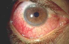
Hemorrhage in the eye
The content of the article:
- Causes of bleeding in the eye
- Forms of the disease
- Stages
- Symptoms
- Diagnostics
- Treatment of bleeding in the eye
- Possible complications and consequences
- Forecast
Hemorrhage in the eye, or hemophthalmos, is a common pathology that can occur both in everyday life and during medical procedures. Sometimes this condition is a complication of various somatic diseases.

Hemorrhage in the eye usually presents with only visual discomfort
Hemophthalmus is the presence of free blood in the vitreous body (vitreum). This structure of the eyeball is a jelly-like substance located between the retina, lens and ciliary body and occupies approximately 70% of the volume of the entire organ.
The soaking of the vitreous with blood is visually determined for several days, after which it is gradually eliminated, which is associated with the breakdown of hemoglobin, which has a characteristic red color. In the course of the destruction of hemoglobin, hemosiderin is formed - an iron-containing pigment that can have a toxic effect on the structural elements of the eyeball.
In addition to local intoxication with hemosiderin, a consequence of hemorrhage in the eye can be the formation of connective tissue cords (moorings) extending to the retina. Under certain circumstances, the presence of such adhesions is one of the conditions for traction (caused by excessive tension) retinal detachment.
Thus, despite the seeming simplicity of the pathological condition and its rather rapid resolution, hemorrhage in the eye can become a prerequisite for serious ophthalmic problems.
Causes of bleeding in the eye
The range of causative factors that can provoke hemorrhage in the eye is quite wide:
- blunt injury, contusion of the eyeball with traumatic rupture of blood vessels (hemorrhage in the eye after a blow);
- penetrating eye injury;
- rhegmatogenous retinal detachment (caused by micro-ruptures of the inner lining of the eye with subsequent penetration of fluid under it);
- surgical intervention;
- atherosclerotic vascular disease;
- vasculitis;
- autoimmune diseases;
- proliferative retinopathy;
- retinal vein occlusion;
- diseases of the coagulation system (coagulopathy);
- rupture of blood vessels against the background of arterial hypertension;
- retinal angiopathy on the background of diabetes mellitus;
- neoplasms of the choroid;
- sickle cell anemia; and etc.
The cause of hemorrhage in the eye can also be the outflow of free blood under the arachnoid membrane of the brain - Terson's syndrome. In this case, a sharp abrupt increase in intracranial pressure leads to an increase in pressure in the retinal vasculature, provoking its damage.
Forms of the disease
There are several forms of hemorrhage in the eye, depending on the amount of damage to the vitreous body:
- partial hemophthalmus - less than 30% of the volume of the vitreous body is soaked in blood;
- subtotal - up to 75% of the vitreum mass is involved in the pathological process;
- total - damage to more than 75% of the vitreous body.

Hemophthalmos, or bleeding in the eye
In accordance with the localization within the vitreous body, hemophthalmos is classified as follows:
- retrolental - hemorrhage is located between the anterior hyaloid membrane and the posterior capsule of the lens;
- paracillary - surrounds the rear of the lens periphery;
- preretinal - between the posterior hyaloid membrane and the retina;
- epimacular - between the posterior hyaloid membrane and the retina in the macular region;
- intravitreal - actually in the vitreum cavity;
- combined - formed by the involvement of several zones of the vitreous body.
Hemorrhage in the eye can be either unilateral or bilateral.
Stages
Hemorrhage in the eye as a dynamic process goes through several stages:
- The stage of bleeding itself (lasts from a few seconds to 24 hours), at which free blood flows into the vitreous body and focal violation of its transparency.
- The stage of a fresh hematoma (up to 2 days), when blood clots are organized.
- Toxic-hemolytic stage (from 3 to 10 days), characterized by the breakdown of hemoglobin with the formation of hemosiderin, active phagocytosis and diffuse opacity of vitreum substance.
- The proliferative-dystrophic stage of hemorrhage in the eye can last up to six months, during this period hemosiderotic dystrophy of the internal structures of the eyeball increases, and connective tissue degeneration of the hematoma occurs.
- The stage of intraocular fibrosis (from 6 months and later), which is characterized by the formation of connective tissue adhesions, persistent hemosiderosis and retinal detachment.
Hemorrhage in the eye does not necessarily go through all stages from bleeding to fibrosis. With timely diagnosis and qualified therapy, hemophthalmos is stopped at the toxic-hemolytic stage. Otherwise, as a rule, further development of the pathological process occurs.
Symptoms
The main manifestation of hemorrhage in the eye is visual impairment of various nature:
- stripes, points, shadows moving in the field of view;
- "Fog", "veil" or clouding before the eyes;
- decreased visual acuity, blurred image outlines up to the level of perception "light-shadow";
- photopsies (bright flashes);
- sudden loss of vision;
- improvement of vision in the first minutes after waking up (organization of outflowing blood in the lower part of the eyeball during sleep).

Photopsy (bright flashes in front of the eyes) - one of the signs of bleeding in the eye
Hemorrhage in the eye is usually not accompanied by painful sensations, manifesting itself exclusively as visual discomfort.
Diagnostics
At an early stage of the disease, with extensive lesions, visual diagnosis of hemorrhage does not cause difficulties.
If the patient asks for help a few days after the hemorrhage in the eye or the lesion is local, limited, a number of studies are needed:
- collection of anamnestic data (the development of hemorrhage in the eye after a stroke, the presence of vascular diseases, the coagulation system, episodes of a sharp increase in pressure in the past, etc.);
- ophthalmoscopy;
- measurement of intraocular pressure;
- Ultrasound examination of the eyeball;
- specific chromatic electroretinography.

To diagnose hemorrhage, ultrasound of the eye and other studies are performed
Ultrasound examination is indicated in the presence of clots of organized blood in the thickness of the vitreous body or in case of massive hemorrhage in the eye, when a full examination of the fundus is impossible.
Treatment of bleeding in the eye
The tactics of treating hemorrhage in the eye depends on its volume: hemophthalmos with a small lesion area usually resolves on its own.
In the presence of active complaints, severe concomitant diseases, a significant extent of the lesion, the patient is hospitalized in an ophthalmological hospital, where special therapy is carried out:
- bed rest, rest, cold eyes;
- in the early days - hemostatic therapy;
- if hemostatic pharmacotherapy is ineffective, laser coagulation of retinal vessels is indicated;
- after a stable stop of bleeding, absorbable drugs are prescribed that contribute to the disorganization of the hematoma;
- topical glucocorticosteroid preparations (for the prevention of mooring);
- physiotherapy treatment (electrophoresis, ultrasound, laser therapy).
At the moment, there are no special drugs with proven efficacy in the treatment of bleeding in the eye.

Vitrectomy (microsurgical removal of damaged areas) is used when conservative therapy is ineffective
In case of insufficient effect of conservative treatment of hemorrhage in the eye, microsurgical removal of damaged areas of the vitreous body (vitrectomy) is used.
Possible complications and consequences
Hemorrhage in the eye can have the following complications:
- toxic damage to the retina (hemosiderosis);
- partial or complete loss of vision.
Forecast
The prognosis for hemorrhage in the eye is generally good. There is a tendency for spontaneous resorption of hematoma in the vitreous body even in the absence of treatment (approximately 1% per day).
The prognosis worsens with subtotal or total hemorrhage in the eye, the presence of moles, signs of retinal detachment, and a long history of the disease.
YouTube video related to the article:

Olesya Smolnyakova Therapy, clinical pharmacology and pharmacotherapy About the author
Education: higher, 2004 (GOU VPO "Kursk State Medical University"), specialty "General Medicine", qualification "Doctor". 2008-2012 - Postgraduate student of the Department of Clinical Pharmacology, KSMU, Candidate of Medical Sciences (2013, specialty "Pharmacology, Clinical Pharmacology"). 2014-2015 - professional retraining, specialty "Management in education", FSBEI HPE "KSU".
The information is generalized and provided for informational purposes only. At the first sign of illness, see your doctor. Self-medication is hazardous to health!


