- Author Rachel Wainwright [email protected].
- Public 2023-12-15 07:39.
- Last modified 2025-11-02 20:14.
Intestinal polyps: symptoms, treatment, prognosis and prevention
The content of the article:
-
Place of localization of polyps
- Small intestine
- Colon
- Types of polyps
- The reasons
- Symptoms of polyps in the intestine
- Diagnostics
-
Treatment of polyps in the intestine
- Electrocoagulation
- Enterotomy
- Resection of part of the intestine
- Diet in the postoperative period
- Forecast and prevention
- Video
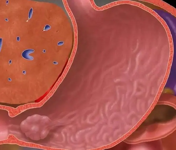
Intestinal polyps are benign neoplasms that can occur anywhere in the intestine. A polyp is a tumor-like outgrowth on a broad base or a thin leg, towering above the mucous membrane into the lumen of a hollow organ (intestine, stomach, uterus, etc.).

Polyps can occur in any part of the intestine
Pathology is a fairly common phenomenon. Most of the growths do not cause any symptoms and are detected by chance during examination. But it must be remembered that in almost 95% of cases, adenomatous and villous polyps become malignant within 5-15 years.
Place of localization of polyps
Small intestine
Rarely enough, formations of this type are found in the small intestine. In the medical literature, isolated cases of the development of neoplasms of such localization are noted. In almost half of patients from this group, polyps are observed in other parts of the gastrointestinal tract (gastrointestinal tract).

The structure of polyps on a broad base
They mainly consist of glandular tissue, but fibromatous and angiomatous ones can occur. Growths on the inner walls of the small intestine have been identified in adults between the ages of 20 and 60.
Localization of polyps in the duodenum is very rare. Almost all patients who consulted a doctor with such a pathology were operated on, since it was suspected that the neoplasm was malignant.
Such outgrowths can be located in the area of the sphincter of Oddi (in patients with cholecystitis or gallstone disease) or near the duodenal bulb (with gastritis with high acidity). The disease occurs in both women and men between the ages of 30 and 60.
Colon
Most often, polyposis formations are located in the large intestine (sigmoid or rectum). They can be either single or multiple. In most cases, they are formed in adolescence, but sometimes they can be detected in children (which may indicate a hereditary predisposition).
Multiple or single growths of such localization are observed in 15% of people after 40 years. In almost 8 out of 10 people, they precede rectal cancer.
Types of polyps
Intestinal neoplasms are classified as follows:
| Type of polyps | Description |
| Adenomatous | The growth is a glandular tissue adenoma. These formations rarely reach large sizes (no more than 1 cm in diameter). In most cases, they have the shape of a mushroom (sometimes they can look like a ball or growth on the mucous membrane), a fairly dense consistency, and a pale pink color. They practically merge with the mucous membrane. Polyps of this type degenerate into a malignant tumor in 1% of cases. |
| Villous |
This is one of the types of adenomatous neoplasms. It is formed from epithelial tissues and can reach large sizes (up to 3 cm). In appearance, they resemble knots on a short, dense leg. Since the villous outgrowths are supplied with a large number of blood vessels, their color can be bright red, as can be seen in the photo. These formations are four times more likely to degenerate into malignant tumors. |
| Glandular villous | They are large lobular growths with a high degree of epithelial dysplasia. The most dangerous are formations, the size of which is more than 1 cm, soft to the touch. They are more likely than others to become malignant. |
| Hyperplastic | They are small growths (up to 0.5 mm in diameter), resembling plaques, located on the mucous membranes of the intestine. In color, they practically merge with the surrounding tissues. They are reborn in a malignant form in very rare cases. |
| Juvenile | In most cases, this type of neoplasm is detected in adolescence. Polyps originate from embryonic tissue debris and are large (up to 5 cm in diameter) round or lobular glossy growths with long legs |
Attention! Photo of shocking content.
Click on the link to view.
The reasons
The causes of the disease are not fully understood and continue to be actively studied.

One of the predisposing factors for the development of pathology is inappropriate nutrition
Factors contributing to the appearance of pathology include:
- hereditary predisposition;
- poor nutrition: eating a lot of fried foods, red meat and animal fats with a minimum amount of vegetables and seafood in the diet;
- chronic somatic diseases;
- intestinal infections;
- chronic constipation;
- alcohol abuse and smoking.
Symptoms of polyps in the intestine
The disease at the initial stages may not manifest itself in any way, proceeding asymptomatically. In some cases, it is possible to identify growths only with a routine examination.
The first signs of polyps in the intestine appear if the formation reaches a large size, begins to ulcerate, or is supplemented by inflammatory processes.
The following symptoms may indicate the presence of masses in the large intestine:
- bleeding. It can occur as a result of ulceration of the outgrowth, torsion of its legs or damage to blood vessels;
- pulling pains: it may hurt in the lower abdomen or in the sacrum;
- frequent urge to empty the bowels;
- mucus in the feces (an indirect sign of villous intestinal polyps);
- pain in the anus;
- alternation of constipation and diarrhea.
Growths located on the walls of the small intestine are very dangerous, as they often degenerate into cancer. They can also cause perforation of the intestinal walls, profuse bleeding or intestinal obstruction.
Signs of a polyp in the small intestine:
- dyspeptic symptoms (belching, nausea, flatulence), usually occur at the initial stage of the disease;
- indomitable vomiting, which occurs in cases where the neoplasm is located in the initial sections of the small intestine;
- cramping abdominal pain;
- bleeding.
In 67% of cases, growths located in the duodenum do not cause any symptoms and cannot be determined. But if the neoplasm reaches a large size, the patient may experience the following symptoms:
- pulling cramping pain near the navel;
- belching a rotten egg;
- feeling of fullness in the stomach;
- frequent nausea.
Diagnostics
Various methods are used to diagnose polyposis (depending on where the growths are located).

To clarify the diagnosis, a colonoscopy may be prescribed.
Diagnostic methods:
- ultrasound examination of the abdominal organs;
- esophagogastroduodenoscopy;
- fluoroscopy;
- colonoscopy;
- CT scan.
It is also necessary to do a fecal occult blood test. A referral for examination can be obtained from a gastroenterologist. In some cases, in order to diagnose the disease, the patient must be admitted to a hospital.
Treatment of polyps in the intestine
The only effective treatment for outgrowths is to remove them. Conservative therapy is carried out only in the presence of diffuse polyposis (when the growths spread to large areas of the intestine) or as a temporary measure before surgery.
Electrocoagulation
If the growth is single, benign and located in the distal colon, it is removed through a colonoscope by electrocoagulation.
Enterotomy
Enterotomy is indicated to eliminate formations in the small intestine or in the duodenal region. The operation is performed under general anesthesia. The surgeon dissects the abdominal wall and removes the bowel loop.

With growths in the small intestine, they are promptly removed
At the next stage, the intestinal wall is dissected in the longitudinal direction and the formation is eliminated. Then the wound is sutured. This operation does not lead to a narrowing of the intestinal lumen, therefore, in the future, the work of the intestine is not disturbed.
Resection of part of the intestine
If there is a suspicion of malignancy of the formation, resection is indicated. A part of the intestine, which has an independent blood supplying mesenteric branch, is removed. After such an operation, the patient may have digestive problems.
Diet in the postoperative period
In order to speed up healing and prevent the formation of new growths, the patient should follow a diet after surgery. He is prohibited from eating spicy, salty and sour foods. It is also necessary to give up fried and fatty foods. The patient needs to reduce the amount of salt in the diet as much as possible.

In the postoperative period, you must follow a diet
Doctors recommend that you eat often (every 2-3 hours) in small portions. Dishes should be at room temperature. They are prepared by boiling, baking or steaming. The consistency of the dishes should be soft, they must first be crushed by rubbing through a sieve or using a blender.
Particular attention should be paid to fluid intake. You need to drink up to two liters of pure non-carbonated water or weak black tea per day. It is worth giving up the use of carbonated drinks and alcohol.
Forecast and prevention
Can a polyp in the intestine disappear on its own? No, such neoplasms do not dissolve, they must be removed surgically.
The prognosis of the disease is favorable if the formation is detected and eliminated on time. The longer a growth lasts, the more likely it is to turn into a malignant tumor.
In order to prevent the development of the disease, it is necessary:
- eat right: give up the use of fatty and fried foods, alcohol, carbonated drinks, introduce vegetables, fruits, seafood into the diet;
- get rid of bad habits;
- effectively and timely treat constipation;
- lead a healthy lifestyle, play sports, walk in the fresh air.
Especially carefully you need to monitor your health for people who are at risk of developing pathology. If intestinal lesions are found in close blood relatives, it is necessary to conduct regular examinations.
Video
We offer for viewing a video on the topic of the article.

Anna Kozlova Medical journalist About the author
Education: Rostov State Medical University, specialty "General Medicine".
Found a mistake in the text? Select it and press Ctrl + Enter.


