- Author Rachel Wainwright [email protected].
- Public 2024-01-15 19:51.
- Last modified 2025-11-02 20:14.
Polyps in the rectum: symptoms, treatment, complications
The content of the article:
- Classification
- The reasons
-
Symptoms of polyps in the rectum
- Stool disorder
- Discomfort in the rectal area
- Stomach ache
- Mucus and blood in the stool
- Diagnostics
-
Treatment of polyps in the rectum
- Transanal excision
- Electrocoagulation
- Transanal endoscopic microsurgery
- Resection
- Traditional medicine methods
- Complications
- Video
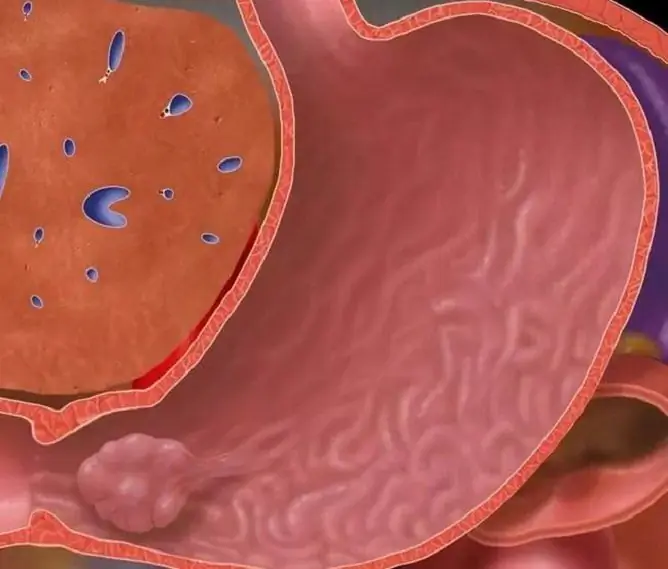
Polyps in the rectum are benign epithelial neoplasms located on the walls of the intestine and growing into its lumen.

Rectal polyps are common
They are found in 7.5% of adult patients during sigmoidoscopy. But doctors believe that there are much more people with this disease, since it is practically asymptomatic. According to some reports, neoplasms in the rectum are found during autopsy in 30% of patients.
Intestinal polyps are considered a rather dangerous precancerous disease, which means that they often degenerate into malignant tumors. They are most common in people who eat large amounts of fatty foods.
Classification
Depending on the histological structure, these neoplasms are classified as follows:
| Type of polyps | Description |
| Glandular | Fibrous rectal polyps develop from glandular tissue and are seen in about 20% of patients. In most cases, they look like a mushroom on a wide leg, but they can also have a branched or spherical shape. |
| Villous (adenomatous) |
This type of growth is also formed from epithelial tissue. They are knots on short wide legs or spread along the walls of the rectum. The villous (fleecy) polyps are rich in blood vessels, therefore they have a bright red color. The size of these formations can reach 3 cm. They often ulcerate and bleed. In 40% of cases, these growths are malignant. |
| Hyperplastic | They are small cysts based on tubular depressions of the intestinal epithelium. These are small neoplasms, the size of which does not exceed 0.5 cm. They have a soft consistency and rise slightly above the surface of the mucous membrane, therefore the disease in most cases is asymptomatic |
Fibrous polyps are quite dense and practically do not differ in color from the mucous membrane. They can reach 2-3 centimeters in diameter. Such neoplasms practically do not bleed and ulcers do not appear on their surface, but in some cases they can degenerate into a malignant tumor.

Neoplasms can have a different histological structure.
Depending on the number of neoplasms, they are classified as follows:
- diffuse: their occurrence is observed with familial polyposis, they are almost impossible to count;
- single: most often it is one large growth;
- multiple: usually polyps grow in groups (in some cases, chaotically).
The reasons
The reasons for the appearance of such neoplasms include:
- chronic bowel disease (proctosigmoiditis, colitis, ulcerative colitis). These pathologies cause degenerative changes in the rectal mucosa, which leads to the formation of polyps;
- acute infectious diseases (salmonellosis, dysentery, rotavirus infection). If they cannot be stopped in the acute period, then structural changes occur in the mucous membrane and the integrity of cellular structures is disrupted, which later becomes a prerequisite for the formation of growths;
- hypodynamia. A sedentary lifestyle leads to congestion, as a result of which the outflow of lymphatic fluid and venous blood is disrupted and edema occurs. This is all aggravated by constipation and forms changes in the rectum for the subsequent formation of neoplasms;
- improper nutrition. Often, neoplasms in the intestines are formed with the frequent use of fatty foods and fast food. The consumption of such food becomes the cause of indigestion and has a negative effect on the mucous membrane;
- hormonal disorders. They occur as a result of endocrine diseases or during menopause in women.
Symptoms of polyps in the rectum
Small formations do not cause any unpleasant symptoms in the patient; they can be detected during examination, which is carried out to diagnose other pathologies. A growth that has reached a large size can manifest itself. In this case, there are signs that are characteristic of other intestinal pathologies.
Stool disorder
This problem appears already at an early stage of the disease. A person has long-term constipation, since a polyp growing into the intestinal lumen prevents the release of feces.
Initially, constipation is rare and is followed by diarrhea. Loose stools become the result of irritation of the mucous membrane.

One of the symptoms of pathology is stool disorder.
Discomfort in the rectal area
With the growth of tissues to medium or large sizes, the formation begins to press on the intestinal wall. Its cavity gradually narrows, and the person begins to feel discomfort in the rectum or on the side of the pubis. Initially, this feeling occurs periodically with the movement of peristaltic waves in the intestine.
If the neoplasms reach large sizes, while a person suffers from constipation, then he experiences discomfort constantly.
Stomach ache
Pain in the lower abdomen is referred to as late symptoms indicating the presence of pathology. Pain occurs when a mass grows substantially and fills the intestinal lumen, which in turn causes constipation.
Mucus and blood in the stool
The presence of blood and mucus in the stool is one of the most common signs of pathology. The reason for this is the hypersecretion of the glands of the mucous membrane. They produce mucus, which moisturizes the rectum and facilitates the movement of feces.
The growth, which is located on the mucous membrane, is an irritating factor and provokes excessive secretion of mucus that accumulates in the intestines. If it is not excreted as a result of constipation for a long time, then it becomes a breeding ground for pathogenic bacteria. Therefore, during bowel movements, mucopurulent discharge may be observed.
Diagnostics
The following methods are used in the diagnosis of the disease:
| Diagnostic methods | Description |
| Finger examination | This is a mandatory primary diagnostic method that allows you to study the structure of tissues in the area of the anus at a distance of about 10 cm. The doctor assesses the condition of the sphincters, the patency of the anal canal, identifies formations and determines the elasticity and mobility of the mucous membrane. Also, during the examination, the specialist detects the presence of blood or mucus. |
| Rectoromanoscopy | The large intestine is examined using a sigmoidoscope (a hollow endoscopic tube equipped with a video camera). The device is inserted through the anus and the folds of the rectum are straightened with air. This method allows you to assess the condition of the mucous membrane, as well as identify pathological changes. If a growth is detected, a biopsy is done (tissue is taken for research) |
| Bowel X-ray | If the diagnosis of neoplasms is difficult, a contrast agent that absorbs X-rays is injected into the intestinal cavity. After filling the intestinal sections, overview and sighting pictures are taken. In the photo, you can identify a growth |
Treatment of polyps in the rectum
Treatment of polyps in the rectum without surgery is not carried out; they are eliminated during surgery. There are different methods of removing such neoplasms.

The method of diagnosis and removal of polyps is determined individually
Transanal excision
This method is used to eliminate polyps that are located near the anus (no more than 10 cm). Before the procedure, the intestines are cleaned with an enema.
The operation is performed under local anesthesia. With the help of a rectal mirror, the anus is expanded. Then the formation is removed, sutures are applied or the vessels are electrocoagulated. At the next stage, the wound is treated with an antiseptic. A tampon soaked in balsamic liniment according to Vishnevsky is introduced into the rectum.
A control examination is carried out two months later. The main disadvantage of this method is the risk of bleeding.
Electrocoagulation
Electrocoagulation is performed if the patient has a single growth up to 3 cm in size, localized at a distance of 10 to 30 cm from the anus.

Electrocoagulation is indicated in cases of a single build-up
The procedure is carried out in the same way as sigmoidoscopy. Pre-purification of the intestines. Then the sigmoidoscope is inserted into the anus and the intestinal walls are examined. After visualization of polyps, a diathermic loop is introduced, with which the pedicle of the formation is captured.
At the next stage, a current is applied to the loop, after which the neoplasm is pulled out. If the build-up is small (up to 0.3 cm), then it is removed by a single touch, as a result of which it is burned. A complication of this procedure can be a perforation of the intestinal wall.
Transanal endoscopic microsurgery
This is a modern effective method that allows you to eliminate polyps in any part of the rectum. Manipulation is performed using a surgical proctoscope. It is injected into the rectal cavity, then carbon dioxide is supplied, which expands the lumen.

Transanal endoscopic microsurgery is most effective for sessile adenomatous polyps
The video camera allows you to detect neoplasms and transmits the image to the screen. Using special instruments, the polyp is excised, and bleeding is eliminated by coagulation. Postoperative complications occur in extremely rare cases (in about 1% of patients).
In comparison with known methods of local removal of rectal growths, transanal endoscopic microsurgery has the following advantages:
- precise excision (thanks to visual control within the muscle layer);
- provision of hemostasis.
The use of this method is most justified when it is necessary to remove adenomatous polyps on a broad basis. Endoscopic microsurgery can be combined with abdominal surgery for synchronous tumor lesions of the colon and rectum.
Resection
This is a radical method that is used when a malignant neoplasm is suspected. The operation is performed under general anesthesia. Initially, an incision is made in the abdominal wall, and in the future, part of the rectum is removed along with the polyp.
If the tumor is malignant, then the rectum is removed completely. In the presence of metastases, lymphatic vessels are eliminated.
Traditional medicine methods
In folk medicine, celandine is used to treat the disease. The sap of this plant contains substances that affect tumors. To prepare the product, a teaspoon of celandine juice is dissolved in 1 liter of warm boiled water. Dry celandine can also be used: one teaspoon of raw materials is poured into 300 ml of water and boiled in a water bath for 20 minutes.
The solution is applied rectally: it is injected into the rectum for 20-30 minutes using a combined heating pad or syringe.
It is necessary to take into account the lack of an evidence base for traditional medicine for neoplasms in the intestine and the existing high risk of complications during inadequate therapy.
Complications
If the detected neoplasms are not removed in time, the following complications may develop:
- malignancy of education (degeneration into a cancerous tumor);
- inflammatory process in the intestine (enterocolitis);
- the formation of fecal stones;
- anemia;
- intestinal obstruction.
Polyps in the rectum are a rather serious pathology, therefore, if you suspect their presence, first of all, you should consult a doctor.
Video
We offer for viewing a video on the topic of the article.

Anna Kozlova Medical journalist About the author
Education: Rostov State Medical University, specialty "General Medicine".
Found a mistake in the text? Select it and press Ctrl + Enter.


