- Author Rachel Wainwright wainwright@abchealthonline.com.
- Public 2023-12-15 07:39.
- Last modified 2025-11-02 20:14.
Umbilical hernia in children
The content of the article:
- Causes and risk factors
- Disease types
-
Symptoms of an umbilical hernia in children
- Embryonic umbilical hernia
- Postnatal umbilical hernia in children
- Diagnostics
- Umbilical hernia treatment in children
- Potential consequences and complications
- Forecast
- Prevention
Umbilical hernia in children is a variant of the hernia of the anterior abdominal wall, in which the internal organs protrude through the expanded umbilical ring. Umbilical hernia in children is very common. The disease is diagnosed in 30-35% of premature babies and 20% of babies born on time. In the structure of the total number of hernias in children (white line of the abdomen, ventral, femoral, inguinal), the share of umbilical hernias is 12-15%. Most often, the disease is diagnosed in girls under 10 years old.

Umbilical hernia is diagnosed more often in girls than in boys
Causes and risk factors
In newborns, after the umbilical cord has fallen off, the umbilical ring closes and gradually becomes invaded (obliterated) with scar connective tissue. Usually, the lower part of the umbilical ring, which contains the umbilical arteries and the urinary duct, contracts better. The upper part, containing the umbilical vein, contracts weakly, since it does not have a muscular membrane.
In the process of strengthening the umbilical ring, the muscles of the anterior abdominal wall, which contribute to its additional tightening, are of no small importance. With a weak tone of the abdominal muscles until the complete obliteration of the umbilical ring, an increase in intra-abdominal pressure when coughing, sneezing or straining contributes to the exit into the umbilical space of the intestinal loops, omentum, and peritoneum. Thus, the formation of an umbilical hernia in children occurs due to the weakness of the peritoneal fascia with incomplete infection of the umbilical ring. The hernial sac with an umbilical hernia in children usually includes loops of the small intestine and an omentum.

Umbilical hernia in children may occur due to incomplete infection of the umbilical canal
One of the main risk factors for the development of an umbilical hernia in children is a hereditary predisposition. It is known that if one of the parents had a pathology, then the probability of its occurrence in a child is 70%.
Also, the risk factors for the formation of a hernial protrusion in a child include diseases and conditions in which intra-abdominal pressure increases, since straining and coughing lead to an increase in the protrusion of the peritoneum and an even greater stretching of the umbilical ring.
These conditions include:
- phimosis;
- chronic constipation;
- lactase deficiency;
- dysbiosis;
- dysentery;
- pneumonia;
- bronchitis;
- whooping cough.
According to statistics, an umbilical hernia is most often observed in prematurely born children, as well as in babies suffering from diseases, against which there is a decrease in the tone of the muscles of the anterior abdominal wall:
- ascites;
- rickets;
- hypotrophy;
- congenital hypothyroidism;
- Down syndrome.
Disease types
Depending on the time of formation and features of the anatomical structure, there are:
- hernia of the umbilical cord, or embryonic (true and false);
- postnatal umbilical hernia.
Symptoms of an umbilical hernia in children
Each form of the disease has its own clinical symptoms, as well as indications for surgical intervention.
Embryonic umbilical hernia
Embryonic umbilical hernias, both true and false, are formed during the period of intrauterine development of the fetus.
A false embryonic hernia is an eventration (prolapse) of the abdominal organs resulting from the underdevelopment of the anterior abdominal wall. This type of umbilical hernia is very rare, about 2-3 cases per 7,000 newborns.

Embryonic umbilical hernias in children occur due to underdevelopment of the anterior abdominal wall
False embryonic umbilical hernias in children are usually combined with other malformations:
- atresia of the anus;
- urachus cyst;
- Meckel's diverticulum;
- congenital intestinal obstruction;
- clefts of the face ("cleft palate", "cleft lip");
- ectopia of the bladder;
- underdevelopment of the pubic articulation;
- congenital heart defects;
- defects in the development of the diaphragm;
- splitting of the sternum.
When examining a newborn, the liver and loops of the small intestine, located outside the abdominal cavity, are visible, covered with a thin translucent membrane. At the time of the passage of the fetus through the birth canal of the mother or in the first hours of the life of the newborn, this membrane can rupture and then the intestinal loops come out.
True embryonic hernia of the umbilical cord (omphalocele or embryonic hernia in children) is formed as a result of abnormal development of the peritoneum at the twelfth week of intrauterine development of the fetus. They occur with a frequency of 1 in 4,000 deliveries.
The hernial sac of embryonic hernias has three layers formed by the peritoneum, vartan jelly and amnion. The protrusion can have different sizes, ranging from 1-2 cm in diameter to 10 cm or more. Germ hernias usually contain part of the liver and intestinal loops in their sac. When crying and straining the child, the protrusion increases markedly.
Postnatal umbilical hernia in children
The formation of postnatal umbilical hernias occurs after birth and clinically they usually begin to manifest themselves in 2-3 months of a child's life.
The leading symptom of an umbilical hernia in children is the appearance in the navel area of a small swelling that has an oval or round shape. When the child cries and strains, this swelling increases.
For most children, an umbilical hernia does not cause any discomfort or anxiety. Only with the progression of the disease and the achievement of a significant hernial protrusion, the child may experience soreness in the umbilical region, constipation, nausea, and cramping abdominal pain.

The main symptom of an umbilical hernia in children is swelling in the navel.
Umbilical hernias in children are infringed rather rarely. If there is a strangulation (infringement, compression) of the intestinal area, then the hernial protrusion becomes irreducible. In this case, a symptom complex of an acute abdomen develops:
- severe cramping pain in the abdomen;
- delay in stool and gas discharge;
- severe nausea and repeated vomiting;
- protective tension of the muscles of the anterior abdominal wall (board-shaped abdomen);
- positive symptom of Shchetkin - Blumberg.
Diagnostics
Diagnosis of an umbilical hernia in children is usually straightforward. On palpation of the anterior abdominal wall, an enlarged umbilical ring is determined. If you raise the child's head and torso, then the hernial protrusion and the area of divergence of the rectus abdominis muscles become noticeable.
If there is a question about the need for surgical removal of an umbilical hernia in children, then a number of instrumental studies are additionally carried out:
- herniography (X-ray examination of the hernial sac with the introduction of a contrast agent into it);
- X-ray of the gastrointestinal tract organs using barium sulfate;
- survey radiography of the abdominal cavity;
- Ultrasound of the abdominal and pelvic organs.

Umbilical hernia on computed tomography
Diagnosis of embryonic umbilical hernias in most cases is carried out even in the antenatal period during ultrasound examination of the fetus.
Umbilical hernia treatment in children
Postnatal umbilical hernias in children are prone to self-healing, so expectant tactics are justified in this case. To strengthen the abdominal muscles, it is recommended to lay the child on his stomach more often, swimming in the pool, physical therapy, massage are also shown.
In some cases, the doctor may prescribe a conservative treatment, which consists in wearing a bandage or applying an adhesive bandage, which mechanically closes the defect in the umbilical ring and thereby prevents its further expansion.

With a postnatal umbilical hernia, doctors recommend placing the baby on the tummy more often
Postnatal umbilical hernias go away on their own by about seven years, that is, by the time the child's abdominal wall gets stronger enough and the umbilical ring is completely closed with connective tissue fibers.
The indications for umbilical hernia surgery in children are:
- preservation of hernial protrusion in children over 10 years old;
- infringement of a hernia;
- digestive disorders;
- significant size of the hernial protrusion.
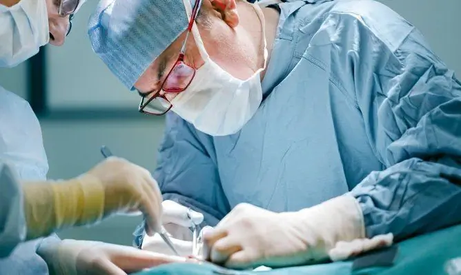
During the operation, the surgeon returns the fallen out internal organs to the abdominal cavity, excises the hernial sac, and then performs plastic surgery (suturing and strengthening) of the area of the hernial orifice, that is, the expanded umbilical ring. The operation usually takes 30-40 minutes. If the child is in good condition, he is discharged from the hospital the very next day.
In case of infringement of an umbilical hernia in children, a necrotic section of the intestine is resected (removed) with subsequent restoration of its integrity (anastomosis is performed using the “end to end” or “end to side” method). Then the hernial sac is excised and plastic of the hernial orifice is performed, the abdominal cavity is drained if necessary.
With an embryonic hernia, surgical intervention is indicated in the first hours or days of a newborn's life.
Potential consequences and complications
The most dangerous complication of embryonic hernia of the umbilical cord is rupture of the membranes that form the hernial sac. As a result, an infection penetrates into the abdominal cavity, leading to the development of diffuse peritonitis.
Infringement of postnatal umbilical hernia in children is accompanied by necrosis of the intestinal area and the development of mechanical intestinal obstruction.
Forecast
The prognosis for embryonic umbilical hernias, especially when combined with other malformations, is unfavorable. The probability of death in this pathology is 30-60%.
Postnatal umbilical hernias are favorable. As a rule, they do not cause discomfort to children and in most cases they pass on their own by the older preschool age. With a significant size of the hernia, hernia repair is performed. After healing, recurrence of an umbilical hernia in children is extremely rare.
Prevention
To prevent the formation of umbilical hernias in children, measures are recommended to strengthen the muscles of the anterior abdominal wall (laying on the stomach, exercise therapy, massage, swimming). In addition, it is necessary to timely identify and actively treat diseases in which there is a weakening of the muscles of the anterior abdominal wall or an increase in intra-abdominal pressure (rickets, pneumonia, whooping cough).
YouTube video related to the article:

Elena Minkina Doctor anesthesiologist-resuscitator About the author
Education: graduated from the Tashkent State Medical Institute, specializing in general medicine in 1991. Repeatedly passed refresher courses.
Work experience: anesthesiologist-resuscitator of the city maternity complex, resuscitator of the hemodialysis department.
The information is generalized and provided for informational purposes only. At the first sign of illness, see your doctor. Self-medication is hazardous to health!


