- Author Rachel Wainwright wainwright@abchealthonline.com.
- Public 2023-12-15 07:39.
- Last modified 2025-11-02 20:14.
Ovarian retention cyst
The content of the article:
- Follicular cyst
- Corpus luteum cyst
- Paraovarian cyst
- Endometrioid cyst
- Video
Retention ovarian cysts are a collective term that combines several types of bulky tumor-like neoplasms. They make up most of the ovarian formations and are divided into the following types:
- follicular (more than 85%);
- corpus luteum cysts (5-7%);
- paraovarian (10%);
- endometrioid (10-15%).
In one group, they are united by the absence of cell proliferation (i.e., uncontrolled division, as in malignant tumors) and the presence of a cavity that is filled with serous or hemorrhagic fluid.
Each of the pathologies occurs in a certain phase of the menstrual cycle - follicular, ovulatory or luteal. Some, such as a corpus luteum cyst, have a clear relationship with pregnancy. They can develop due to disruption of the hormonal system.
Most often occur in women of young and middle age.
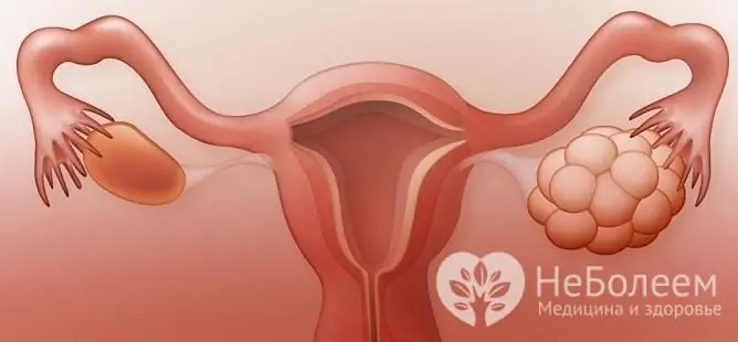
There are several types of ovarian retention cysts
Follicular cyst
This type refers to temporary or functional formations, exists for 1-2 menstrual cycles. The cause of the occurrence is associated with a violation of the ovulation mechanism - the follicle is filled with serous contents instead of bursting with the release of the egg.
The main symptoms are:
- formations up to 5-7 cm in size do not appear clinically;
- sometimes there is a violation of menstruation (delay, soreness, profuse bleeding);
- pain in the lower abdomen (mild, localized on one side).
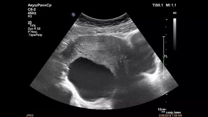
Symptoms appear when complications develop. These include torsion and rupture of the cyst with the development of a symptom complex of an acute abdomen. In this case, the cystic formation mimics other diseases of the abdominal organs. An emergency hospitalization in a surgical hospital and surgical intervention is required. The diagnosis is clarified with the help of a physical examination and instrumental diagnostic data (ultrasound).
The clinical picture of complications:
- Sharp intense pain in the lower abdomen. More often there is a retention cyst of the right ovary, so the pain is usually localized to the right or shifts somewhat towards the bosom.
- Nausea, vomiting. They are a manifestation of the intoxication syndrome. With frequent vomiting, it is necessary to carry out differential diagnosis with rotavirus infections.
- Constipation or loose stools. The symptom will depend on the location of the cystic formation and the degree of intestinal involvement in the inflammatory process.
- The temperature rises to 39 ° C. It is also a manifestation of intoxication syndrome and is present in 60-70% of women.
- Vaginal discharge.
- Tachycardia and decreased blood pressure. The second symptom is especially well manifested in the presence of internal bleeding (when the ruptured follicle was located next to large vessels). Tachycardia acts simultaneously as a symptom of intoxication and centralization of blood circulation.
Management of patients with a confirmed ovarian cyst:
| The form | Time | Treatment |
| Uncomplicated | 6-8 weeks (1-2 phases of the menstrual cycle) |
Shown observation, taking anti-inflammatory drugs and oral contraceptives. Tactics change in the following cases: · Pathological growth (increase in a short time by more than 2 times); · Change of boundaries according to ultrasound data (uneven, blurred); · Change in the content (inhomogeneous, signs of additional inclusions); · Appearance of additional cameras. If one of the presented conditions is present, diagnostic laparoscopy is indicated to clarify treatment options (conservative or surgical). It is also used for differential diagnosis with malignant tumors. |
| Complicated | Signs of a cyst persist for more than 8 weeks or a clinic of torsion and rupture |
Surgical treatment. Management tactics: Hospitalization (in severe conditions, first in the intensive care unit); Restoration of normal parameters of red blood in case of suspected bleeding (hemoglobin, erythrocytes); Restoration of normal blood coagulation values in case of suspected bleeding (coagulogram indicators); · If necessary, bringing the saturation up to 95-98%; · Diagnostic laparoscopy (clarification of the diagnosis); · Laparoscopy or laparotomy, depending on the specific situation and the required amount of intervention. After surgery, the use of folic acid and vitamin complexes, as well as oral contraceptives is shown to restore the female cycle. |
Possible operational tactics:
- unilateral ovariectomy (complete removal of the ovary);
- ovarian resection (removal of only a part of the ovary with a ruptured neoplasm).
The prognosis is favorable even after surgery.
Corpus luteum cyst
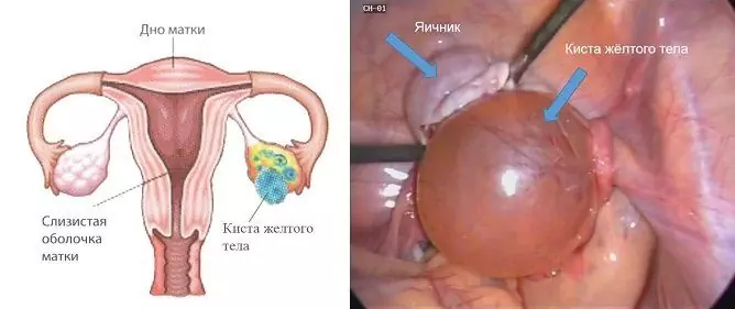
It is found relatively less frequently than other formations. The corpus luteum arises in the ovary under the action of the pituitary hormone, belongs to the temporary endocrine glands and produces progesterone, which ensures the introduction and fixation of the ovum into the uterus. The time of occurrence is the ovulatory phase. It has a rounded shape and a yellowish tint, which gives a specific lipochromic enzyme in the walls.
Disease symptoms:
- clinically not manifested in 90% of women;
- rarely causes menstrual irregularities;
- can cause problems with conception and bearing.
Frequent complications include bleeding into the cavity of the formation, which occurs due to a good blood supply. Sometimes there are ruptures, their clinical symptoms are similar to follicular cysts (symptoms of an acute abdomen), except that pain appears more often on the left (more often there is a retention cyst of the left ovary).
Management tactics:
- Observation of 1-3 phases of the menstrual cycle. Spontaneous resolution in most cases. Treatment should be started under the same criteria as for follicular cystic formations (growth, boundaries, content).
- Treatment of an ovarian retention cyst by surgery occurs in the presence of complications. The tactics are the same as for follicular formations. Feature - laparotomy is rarely used.
- Removal (enucleation) of cystic formation within healthy tissues is performed using laparoscopy.
Paraovarian cyst
Cystic neoplasm that does not belong to the classic lesions of the ovary. It is formed from the embryonic rudiment, which is located in the wide ligament of the uterus between the uterus, ovary and fallopian tube. In rare cases, it gets drunk with the ovary, disrupting its function.
Clinical picture:
- Most often, clinical manifestations are absent.
- Since the cysts grow to large sizes (more than 20 cm), sometimes there are pulling pains in the lower abdomen. On examination, there is a slight asymmetry of the lower abdomen.
- On palpation, a tightly elastic formation can be found in the right or left iliac region.
- Violation of the menstrual cycle and problems with conception (high risk of infertility with large cysts and prolonged absence of treatment).
The clinical picture of complications:
- Torsion and rupture - acute abdomen clinic.
- Adhesion process in the abdominal cavity - depending on the location, symptoms of intestinal obstruction or symptoms of obstruction of the fallopian tubes occur.
- Hemorrhage and suppuration of the cyst. Do not manifest clinically until the rupture and release of the contents of the cyst into the pelvic cavity.
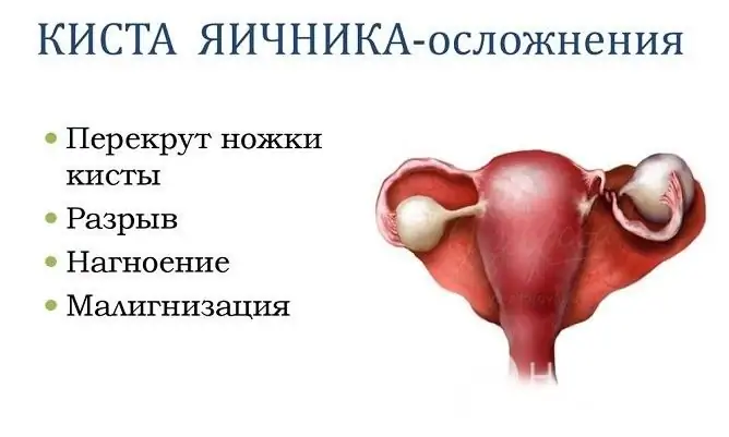
Retention ovarian cysts usually only need to be surgically removed if complications develop
Management tactics:
- Expectant tactics for three menstrual cycles (ultrasound control once every 3-4 weeks). Simultaneous administration of monophasic combined oral contraceptives and a course of anti-inflammatory therapy.
- Planned treatment in the absence of regression after the 3rd cycle of menstruation.
- Emergency hospitalization in the presence of complications.
Two types of access are used during operations:
- laparoscopy - hammer-traumatic, highly effective, allows you to operate on complications;
- laparotomy - wide access, good sanitation of the abdominal cavity (used for cysts larger than 10 cm).
Surgical options:
- unilateral tubo-ovariectomy (removal of the ovary, fallopian tubes);
- enucleation with dissection of the leaf of the broad ligament of the uterus and the intraligamental space (the ovary and fallopian tube are not affected).
The prognosis is relatively favorable.
Endometrioid cyst
It develops due to the cells of the endometrium of the uterus that have migrated into the ovarian cavity. This phenomenon can occur with traumatic injuries of the uterus, recent surgical interventions on the uterus.
Clinical manifestations:
- Initially, there are no clinical manifestations.
- As the cyst progresses, something like erosion (heterotopia) appears on the surface of the cyst, from which the contents of the cyst begin to seep into the abdominal cavity.
- Oiled clinic of the acute abdomen, since the penetration of cystic contents does not occur simultaneously.
- Late adhesive obstruction and, as a result, problems with conception.
- Provided that endometrial cells grow, bloody discharge is possible (the volume of discharge varies).
Complications and their clinic are similar to those in paraovarian cystic formations. Management tactics (depending on the stage of endometriosis):
- Planned surgical treatment (stage 3-4 of endometriosis) - separation of adhesions, removal of the cyst with a capsule (husking). Ovarian resection is extremely rare. After the operation, a course of hormonal therapy is shown for 6 months.
- In the presence of complications, emergency surgery.
The forecast is favorable.
Video
We offer for viewing a video on the topic of the article.

Anna Kozlova Medical journalist About the author
Education: Rostov State Medical University, specialty "General Medicine".
Found a mistake in the text? Select it and press Ctrl + Enter.


