- Author Rachel Wainwright wainwright@abchealthonline.com.
- Public 2023-12-15 07:39.
- Last modified 2025-11-02 20:14.
Parapelvic cyst of the kidney
The content of the article:
- Specifications
- Classification
- Features:
-
The reasons
- Congenital
- Acquired
-
Treatment
- Surgery
- Conservative therapy
- Forecast
- Video
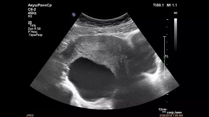
Parapelvic cyst of the kidney refers to benign neoplasms and is one of the forms of a simple solid cyst. It is less common than other forms (parenchymal, subcapsular).
Specifications
- It is localized strictly at the gate of the kidney and does not affect the tissue of the organ itself (pelvis, sinuses).
- More often there is a parapelvic cyst of the left kidney (about 60% of cases).
- The cavity is always filled with fluid (in the presence of a tissue component or other inclusions, differential diagnosis with malignant tumors is necessary).
- Rarely reaches large sizes.
- Symptoms occur with small cysts, since the lumen of the renal pelvis becomes obstructed, which causes a violation of the outflow of urine.
- There is a high risk of developing acute renal failure.
- Rarely affects both kidneys (all types of simple solitary cysts are characterized by a one-sided manifestation of the disease).
Classification
The classification is given for the purpose of differential diagnosis of closely related formations.
| View | Localization | Features: |
| Pyelogenic |
It is located in the parenchyma of the kidney and has a communication with the lumen of the pelvis or with the calyx. Occurs in newborns or the elderly |
Often located in the middle calyx, less often in one of the poles of the kidney |
| Parapelvic | Located at the gate of the kidney or has a sinus location | Has no communication with the pelvis |
| Peripelvic (calyx cyst, calyx diverticulum). |
Subspecies: Intralocal type - located directly in the pelvis and often soldered to its walls; Intramural - located directly in the wall of the pelvis, in the muscle layer; Extrapelvic - located outside the pelvis and has exophytic growth |
Features depend on the subspecies |
Features:
The structure, features, degree of malignancy and appearance (photo) are assessed according to instrumental diagnostics using the Bosniak scale.

The degree of malignancy is assessed using the Bosniak scale
| A type | Features: |
| Bosniak-I | This category includes a simple parapelvic cystic cavity with typical manifestations: small (up to several centimeters), thin-walled, without septa and calcifications. Filled with serous clear fluid. Does not retain contrast and has a smooth, rounded shape. As a treatment, observation with periodic control of ultrasound is indicated (every six months). |
| Bosniak-II |
Neoplasms are somewhat more complex in structure. Walls with single calcifications. 1-2 partitions appear. The outline is still clean and crisp. The contrast does not accumulate (excretory function is normal). There are no signs of malignancy. No treatment is required, only strict control. |
| Bosniak-IIF | The number of partitions is increasing. The walls thicken. The contrast is not displayed completely (by 90%). No curing is required, strict control is needed. |
| Bosniak-III | The wall is dense, there are multiple calcifications in the walls. Multi-chamber. There are more than 5 partitions in the cavity. The contrast is displayed only by 50%. The content is represented by a serous, hemorrhagic and protein component. Surgical treatment is required. |
| Bosniak-IV | Presented by malignant forms (malignancy 100%). An operation is required as planned. |
In fact, this classification shows the progression of a benign cystic neoplasm to malignant tumors (the likelihood of malignancy increases). Two diagnostic methods are used for assessment: CT and ultrasound.
The reasons
The causes are divided into congenital and acquired.
Congenital
Congenital malformation of the renal tissue, which is associated with blockage of the lymphatic vessels near the remnant of the mesonephros (wolffian duct). In some cases, there is atresia of the lymphatic ducts at the site of the kidney. In both cases, lymph accumulates in the sinuses and leads to their expansion and deformation of the pelvis, calyces. In this case, a typical simple cystic cavity arises, but if it occurs next to the pelvis, it is classified as a parapelvic cyst of the right kidney or left. It is found in the fetus during a routine examination of pregnant women (II, III trimester). No emergency surgery is required. Termination of pregnancy is not indicated.
Acquired
- Inflammatory processes (glomerulonephritis, pyelonephritis). Cystic formations occur as a complication of bacterial or viral kidney damage. Such a manifestation of infectious pathology occurs extremely rarely.
- Urolithiasis disease. Neoplasms can occur due to mechanical damage or obstruction of the lumen of the urinary tract.
- General infectious diseases (kidney tuberculosis). In this case, cystic cavities in the parenchyma often appear, but mycobacteria are capable of infecting any renal structure.
- Benign and malignant tumors of the abdominal cavity and retroperitoneal space (neuroblastoma). Often there is mechanical compression of the renal tissue, less often metastatic lesion.
Treatment
Surgery
The main treatment for all cystic formations is surgery. The specific method is chosen by the doctor depending on the individual characteristics of the organism, the localization of the cyst and the presence of complications (suspected rupture). In addition, the type of cystic neoplasm according to the Bosniak classification must be taken into account.
| Operation type | Complications | Features: |
|
Puncture options: · Aspiration puncture of the cyst; · Cyst aspiration puncture and sclerotherapy; Cyst aspiration puncture, drainage and sclerotherapy |
Rarely observed: · Abscess; Hemorrhage (with the formation of hematomas); · Hematuria; Relapses |
A feature is simplicity in technical implementation. It is performed strictly under ultrasound control and has minimal invasiveness. A special biopsy needle is inserted into the cyst cavity under control. The resulting fluid is sent for histological and cytological examination. Then a sclerosing substance is injected into the cavity and the walls of the cyst are soldered. Drainage is rarely used. A stylet-catheter is used as drainage. This is done for partial sclerosis, since with large cysts, it is impossible to inject the entire sclerosant at once (the risk of kidney rupture). |
|
Radical laparoscopic cyst removal: · Transabdominal access; · Translumbar access; Transthoracic access |
Typical for any laparoscopic surgery (extremely rare): · Damage to internal organs when placing a trocar; · Development of peritonitis with damage to the intestine; · Adhesions in the postoperative period; Bleeding; · With transabdominal access; · Accession of a secondary infection; Gas embolism due to carboxyperitoneum; Pneumothorax and pneumomediastinum with transthoracic access |
At the moment, the most effective method of surgery. The placement of trocars and laparoscopes will depend on the approach chosen. Benefits: · Gives a wide view without the need to expand the surgical wound; · Cosmetic; · Fast postoperative period; · Relatively rare relapses (in comparison with puncture). An especially effective method for superficial cysts and in the absence of large great vessels around the formation. They use less often open access (relatively new method). |
|
Open surgery from the lumbotomy access: · Resection of the kidney area with cystic formation; Nephrectomy (complete removal of the kidney on one side) |
Any abdominal operation is highly invasive, since it is done through a wide access. Complications: · Suppuration; Bleeding; · Ventral hernia; · Repeated surgical interventions; Peritonitis; Retroperitoneal hematoma |
The technique is familiar to all surgeons (does not require special knowledge). Layer-by-layer tissue dissection is performed. The muscles are pushed back in a blunt way. The kidney is examined and further tactics are determined (may differ from the initially chosen one). Removal of the cyst and thorough hemostasis are performed. The kidney tissue is sutured and the clamps are removed from the large vessels. Drainages (2) are installed and the wound is sutured in layers. Has a long postoperative period. |
Conservative therapy
It can be used in the preoperative or postoperative period, depending on the clinical picture. Apply:
- antibiotics - when an infection is attached (Furazidin, Furamag, Cefepim);
- antihypertensive drugs - as a component in the treatment of arterial hypertension (Captopril, Enalapril, Verapamil);
- analgesics and antispasmodics - to relieve pain syndrome (Papaverine, Analgin);
- non-steroidal anti-inflammatory drugs (Diclofenac);
- diuretics - with stagnation of urine in the kidneys and disorders in general tests (Spironolactone).
Forecast
With timely detection and treatment, the outcome is favorable.
Video
We offer for viewing a video on the topic of the article.

Anna Kozlova Medical journalist About the author
Education: Rostov State Medical University, specialty "General Medicine".
Found a mistake in the text? Select it and press Ctrl + Enter.


