- Author Rachel Wainwright [email protected].
- Public 2023-12-15 07:39.
- Last modified 2025-11-02 20:14.
Cyst in the head of a newborn
The content of the article:
- Why is cystic formation formed?
- Types of cystic formations
- How does the disease manifest
- Diagnostic methods
- Consequences and possible complications
-
Treatment methods
- Conservative treatment
- Surgical intervention
- Video
A cyst in the head of a newborn is a rather rare disease that is often diagnosed even during pregnancy. The main question that worries parents is what are the consequences of this disease. The danger depends on the type of education. Some cysts dissolve on their own and do not harm the baby. Others are capable of increasing intracranial pressure, leading to a delay in physical and mental development. In any case, it is necessary to consult a doctor and determine the need for treatment of pathology. Sometimes observation is enough, less often surgery and additional medication are prescribed.

A brain cyst in a newborn baby is not always a dangerous formation
Why is cystic formation formed?
A cyst is a benign neoplasm, which is a cavity with liquid contents inside. It often forms in a child in the womb due to the accumulation of fluid in places where dead brain cells accumulate.
No single cause has been established that would lead to the development of pathology. A number of factors can influence the formation of a neoplasm at once:
- Genetic abnormalities. This is influenced by poor ecology, the use of genetically modified products.
- Inflammatory process - genital herpes, toxoplasmosis.
- Postponed trauma. For example, a head injury during childbirth, a difficult pregnancy.
- Autoimmune processes, when the body perceives its own tissues as foreign and attacks them.
Types of cystic formations
Cystic cavities can form in the white or gray matter of the brain, in the thickness of the meninges. There are three main types: arachnoid, subependymal and vascular plexus cysts.
| View | Localization, features |
| Arachnoid | Arachnoid cysts contain CSF inside. They are located between the arachnoid (arachnoid) membrane and the surface of the brain. Such formations do not dissolve on their own and are dangerous due to possible violations of CSF dynamics. |
| Subependymal | The formation is localized under the ependyma of the ventricles. Ependyma is the thin layer that lines the walls of the ventricles of the brain. Ependyma cells contain cilia that help circulate cerebrospinal fluid. Formation of a cyst under the ependymal ventricles can lead to cerebrospinal fluid hypertension. |
| Choroid plexus cyst |
The most favorable type of pathology. It is often formed during pregnancy and passes without consequences. If it occurs after the birth of a child, in 90% of cases it is associated with herpes or other viral infection. In this case, the prognosis depends on the timeliness of treatment. |
How does the disease manifest
In most cases, the disease is asymptomatic, and is detected during an ultrasound examination (ultrasound). Less often, clinical symptoms occur, which depend on the type of neoplasm, its location and size.
| Symptom group | Explanation |
| Local changes | Examination can reveal bone deformity of the cranial vault, fontanel tension. |
| Increased intracranial pressure |
CSF hypertension can manifest itself in various symptoms: Vomiting not related to food intake; · Periodic convulsions; Fontanel tension; Lethargy, drowsiness. |
| Focal symptoms |
Sometimes there are various focal symptoms: • loss of vision; · Hearing loss; Hemiparesis (muscle weakness of the right or left side of the body); Ataxia. |
The clinical picture is often dominated by cerebral symptoms associated with cerebrospinal fluid hypertension. Focal symptoms occur less frequently, for example, when a cystic formation ruptures. At a later age, the child may experience a delay in physical or mental development.
Diagnostic methods
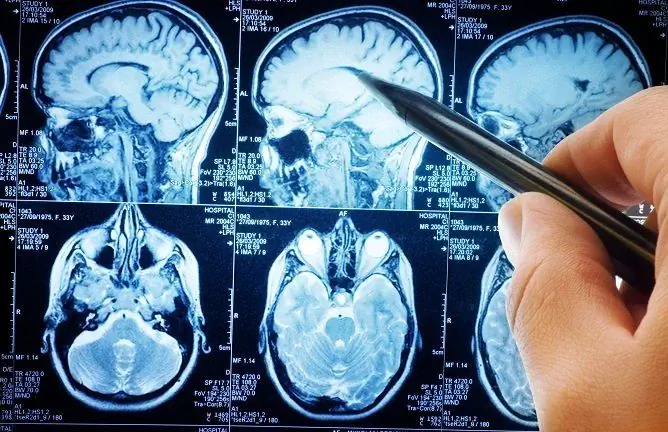
To make a diagnosis, clinical manifestations are not enough, an additional examination is required. In most cases, cystic formations are detected in the fetus even during pregnancy. The main method for diagnosing the disease in newborns is an ultrasound examination of the brain (neurosonography). An ultrasound scan is a safe test that will not harm the baby. Additionally, magnetic resonance imaging (MRI) is prescribed.
| Diagnostic method | Description |
| Brain ultrasound |
Screening diagnostic method. With the help of ultrasound, you can find: · The size of education; · Its localization; · Shape and boundaries; · Communication with the ventricles of the brain. Based on these signs, one can suspect the presence of a cystic cavity and suggest its appearance. However, it is not always possible to make a definitive diagnosis. |
| MRI examination | With the help of MRI, the diagnosis is clarified. This is a more informative and specific diagnostic method that allows you to finally determine the type of formation, its location and size. |
Consequences and possible complications
The prognosis depends on several factors - the type of education, size and location. Small cystic formations often resolve on their own without causing any pathological changes.
The prognosis depends primarily on the size of the cystic formation. When the size is large, there is often an increase in intracranial pressure, which can lead to hydrocephalus. In this case, the prognosis is relatively unfavorable - there may be a delay in mental and physical development, frequent convulsions, and focal symptoms develop less often.
Complications may develop, the most common are:
- infection;
- rupture of the cyst;
- damage to the neurovascular structures;
- hemorrhage.
Treatment methods
Not all formations of the brain are subject to active treatment. Some of them resolve on their own and require only observation. For active treatment, conservative or surgical methods are used.

Sometimes it is enough just to control the cyst with ultrasound; treatment may not be required
Conservative treatment
It is impossible to get rid of cystic formation with the help of medications or folk remedies. But sometimes conservative therapy is still used. It is indicated in cases where the onset of pathology is associated with an inflammatory process. Since viral infection is the most common cause, antiviral drugs may be prescribed. For example, Acyclovir for herpes.
Surgical intervention
The main method of treatment for this pathology is surgery.
What are the indications for surgery:
- hypertensive syndrome;
- the appearance of focal symptoms;
- a progressive increase in the size of the neoplasm;
- development of other complications.
Treatment is carried out by the following methods:
- Liquid shunt operations. Allow to drain the cyst into the subdural space or abdominal cavity.
- Drainage by needle aspiration.
- Excision of the formation using endoscopic techniques.
- Craniotomy with excision of the cystic formation.
When determining the tactics of surgical treatment, the shape, size and localization of the formation are taken into account. It is contraindicated to carry out the operation with an active inflammatory process, decompensation of the child's vital functions.
Video
We offer for viewing a video on the topic of the article.

Anna Kozlova Medical journalist About the author
Education: Rostov State Medical University, specialty "General Medicine".
Found a mistake in the text? Select it and press Ctrl + Enter.


