- Author Rachel Wainwright [email protected].
- Public 2024-01-15 19:51.
- Last modified 2025-11-02 20:14.
Hematoma on the head of a newborn after childbirth: causes, treatment, complications
The content of the article:
- Features:
- Classification
- The reasons
- Characteristics of cephalohematoma
- Therapy
- Complications and outcomes
- Video
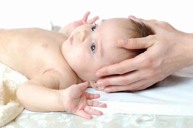
A hematoma on the head of a newborn after childbirth (cephalohematoma) is a visible hemorrhage that occurs immediately after childbirth. It usually appears after the birth tumor has subsided, as a rule - on days 2-3.

Cephalohematoma is one of the birth injuries
The process of delivery is physiological, in which not only the mother, but also the child himself, actively participates. During the passage through the birth canal, the baby performs several biomechanisms that are not easy for him. This is necessary for a less traumatic birth. In addition, for the same purpose, the soft bones of the skull in the place of the seams touch, and sometimes even push against each other.
These circumstances predispose, and in combination with some common causes, can lead to hematoma.
Features:
Often, hemorrhage is localized and limited to the parietal bone, less often to the occipital.

A hematoma forms between the periosteum and the outer surface of the cranial bones
The mechanism of hemorrhage is the rupture of the vessels passing between the periosteum and the outer surface of the cranial bones. This is often due to the displacement of the skin with the periosteum during labor. Blood from damaged vessels seeps between the periosteum and the bone, forming a soft, towering cushion that is visible to the naked eye.
The accumulated blood does not clot for a long time due to the lack of some factors of the neonatal hemocoagulation system.
Classification
Cephalohematoma is classified into three grades, depending on its diameter:
- No more than 4 cm.
- 4.1 to 8 cm.
- More than 8 cm.
Cephalohematoma can be combined with other injuries resulting from childbirth, namely, a fracture of the skull bones, hemorrhage into the subdural space with the formation of a subdural hematoma, hemorrhage into the brain substance, cerebral edema. Pathology may be accompanied by focal and cerebral neurological symptoms.
Areas of localization of cephalohematoma:
- parietal;
- frontal;
- occipital;
- temporal.
The reasons
The appearance of hemorrhage can provoke some factors observed on the part of the mother and / or the fetus.
Common causes include:
- Large fetus: for example, as a consequence of uncompensated diabetes mellitus in the mother.
- Pregnancy lasting more than 40 weeks (post-term): This leads to compaction of the skull bones and inability to configure during passage through the birth canal.
- Congenital malformations of the fetus.
- Pelvic, facial and other types of abnormal presentation and position.
- Narrow pelvis (anatomically or clinically) in the mother.
- Pathology of labor: discoordination, rapid or protracted labor.
- The use of medical instruments to extract the fetal head during childbirth (forceps, vacuum extractor).
- Pregnancy in a woman over 35 years old.
- Umbilical cord entanglement, aspiration of amniotic fluid.

Pathology may be the result of the use of medical instruments for retrieving the fetus during childbirth
Characteristics of cephalohematoma
As a result of the presence of a birth tumor in the first 2-3 days, the hematoma cannot always be immediately recognized. It manifests itself as the birth tumor subsides and can increase for several more days due to the fact that the neonatal coagulation system is not completely formed.
By palpating it, you can feel the presence of fluid under the skin, which, with slight pressure, moves, but does not go beyond one bone.
Differences between cephalohematoma and a generic tumor:
| Differences | Cephalohematoma | Birth tumor |
| Formation mechanism | Formation occurs as a result of hemorrhage | It appears due to swelling of soft tissues, it can be localized not only on the head, but also on other parts of the body (buttocks, thigh, perineum). It depends on which part of the baby's body will first pass through the birth canal. |
| Distribution rate | Hematoma is limited to only one bone of the skull | May extend to multiple bones of the skull |
| Palpation response | With pressure on the hematoma area, fluid rolling is felt | Swelling to the touch dense, doughy |
Therapy
Cephalohematoma does not always require treatment, as it usually resolves on its own within 2-4 weeks.
In the presence of a large hematoma (II-III degree) or the absence of a tendency to regress it, it is necessary to carry out inpatient and / or surgical treatment, taking into account the following principles:
- The child should be under constant supervision by doctors: a pediatric surgeon and a neonatologist, if necessary, a photo is taken for control.
- Conservative treatment consists in the administration of hemostatic drugs (Vicasol, calcium preparations).
- Indication for surgical intervention (puncture and aspiration of hematoma) - cephalohematoma of the III degree.
The duration of inpatient therapy depends on the presence of complications: uncomplicated cephalohematoma is treated from a week to 10 days, complicated - more than a month. Dispensary observation by a pediatric surgeon and neurologist lasts for 1 year.

After inpatient treatment, long-term dispensary observation is necessary.
Cephalohematoma may be the result of diseases of the hemocoagulation system of an infant with a tendency to hypocoagulation (poor blood clotting), for example, in hemophilia, thrombocytopenia, etc. In this case, it is first necessary to treat the underlying disease that caused the formation of hemorrhage.
Complications and outcomes
Complications of cephalohematoma are rare, but in some cases, if treatment is not started in a timely manner, it can lead to serious consequences.
Possible complications:
- calcification of hematoma (ossification), which leads to visible deformations of the bones of the skull;
- yellowness of the skin (due to the destruction of erythrocytes released as a result of vascular damage at the site of hemorrhage);
- a decrease in the amount of hemoglobin and red blood cells (anemia);
- infection of the hematoma with the subsequent formation of a purulent focus.
Attachment of a bacterial infection (infection) with subsequent suppuration can lead to dangerous conditions, therefore, in such cases, it is necessary to puncture the purulent focus, aspiration of the contents, drainage, followed by the application of dressings with antiseptics and antibacterial drugs as soon as possible.
Most often, cephalohematoma passes without a trace and does not require any treatment; in these cases, they speak of a favorable prognosis.
However, sometimes the consequences of a hematoma on a newborn's head can be unfavorable and include the following disorders:
- Delayed mental and speech development of the child.
- Development of infantile cerebral palsy.
- Neurological spectrum disorders.
The best thing a mother can do is to observe the condition of the child and the dynamics of the hematoma. If, 2 weeks after delivery, the site of hemorrhage does not resolve on its own, it is necessary to contact a neonatologist or pediatric surgeon to determine further tactics for managing the baby.
Video
We offer for viewing a video on the topic of the article.

Anna Kozlova Medical journalist About the author
Education: Rostov State Medical University, specialty "General Medicine".
Found a mistake in the text? Select it and press Ctrl + Enter.


