- Author Rachel Wainwright [email protected].
- Public 2024-01-15 19:51.
- Last modified 2025-11-02 20:14.
Hematoma on the head after a blow: features, diagnosis, treatment
The content of the article:
- The causes of the appearance of pathology
-
Types of hematomas
- Epidural hematoma
-
Subdural hematoma
Chronic subdural hematoma
- Symptoms
- Diagnostics
-
Treatment
- Treatment of chronic subdural hematomas
- Home treatment
- Forecast
- Video
A bruised or bruised scalp is a serious and potentially life-threatening condition that often requires immediate treatment. Brain damage can have a lasting impact on the emotional, mental state and health of a person in general.

Most often, a hematoma on the head appears as a result of injury
The brain is a protected organ of the body, but in accidents and accidents, this protection is often insufficient. Intracranial hematoma (hemorrhage) is considered one of the most severe traumatic brain injuries.
The causes of the appearance of pathology
The brain is located inside the cranium, it is enclosed in soft membranes and washed by blood and cerebral fluid. They occupy a volume of 1.5-2 liters. These fluids are incompressible, therefore, if the blood begins to pour into the cranial cavity during a rupture of a vessel or a blow, the brain is displaced - dislocation.
Of the total number of head injuries, hematomas make up no more than 10%. However, they are the ones that give the highest mortality rate. The most serious cases of hospitalization occur in unconscious victims.
In addition to traumatic etiology, hemorrhage can be of a vascular or iatrogenic nature, occur due to hemophilia, craniocerebral imbalances, sepsis, brain tumors, intoxication, etc.
Types of hematomas
The brain, compressed by intracranial hemorrhage, can move, as a rule, into the foramen magnum and the opening of the cerebellar tentorium. At this level in the brain stem is the center of breathing, sensory and motor pathways, as well as a single pathway for the outflow of cerebrospinal fluid from the brain.
The consequence of a violation of the outflow in the cranium will be an increase in the excess volume of cerebrospinal fluid.

There are two types of hematomas on the head
Intracranial hemorrhage is of two types:
| Type of hematoma | Description |
| Epidural | Formed over the dura mater due to arterial bleeding (mainly due to damage to the middle meningeal artery), as a rule, in the place where the blow fell. Characterized by a progressive increase in the size of the lesion |
| Subdural | Formed under the dura mater due to rupture of cortical bridging veins, usually on the side of the skull opposite to the blow |
In some cases, both types of pathology are formed, the symptoms of which may be unexpressed.
Epidural hematoma
This is an accumulation of blood between the inner surface of the skull and the periosteum (the outer layer of the dura mater). In this type of disorder, most often there is a history of trauma and a concomitant fracture of the skull.
Usually the source of bleeding is the rupture of the sharp edges of the fracture of the skull of the branches of the meningeal artery (in most cases, the middle meningeal artery).
Most often, the formations are limited to cranial sutures and have a biconvex shape. They can have a volumetric effect and cause wedging of brain tissue.
In a fairly large number of cases, this type of hemorrhage is unilateral, however, there are bilateral and multiple epidural hemorrhages.
Subdural hematoma
This is an accumulation of blood in the space between the dura mater and the arachnoid. It occurs in any age group of patients:
- children: hemorrhages usually occur as a result of accidental injury, more often they are bilateral; interhemispheric subdural lesions are predominantly observed in children with non-road traffic injuries;
- young people: the most common cause of hemorrhage is road traffic accidents; unilateral hematomas are more common in adult patients;
- elderly: bruising on the head is usually associated with a fall.
Typical sites of localization of hemorrhages are the middle cranial fossa and the frontal-parietal regions.
Chronic subdural hematoma
Chronic subdural hematoma is called a volumetric polyetiological intracranial hemorrhage, which is located under the dura mater, causes general and / or local compression of the brain and has (in contrast to acute and subacute forms) a delimiting capsule.
The capsule usually occurs two weeks after the hemorrhage. These characteristics determine all the features of cerebral pathophysiological reactions, clinical course and therapeutic tactics.
Symptoms
The symptomatology of hemorrhages on the head may not be expressed, most often it manifests itself in the form of a headache. You may also experience such an unpleasant sensation as slight dizziness when changing body position or moving in front of the eyes of transport.

One of the main symptoms of pathology is headache.
Other possible signs of pathology:
- vomiting, nausea;
- drowsiness;
- confusion of consciousness;
- loss of speech or delayed speech;
- weakness in the limbs on one side of the body;
- the difference in the size of the pupils.
Diagnostics
Before starting to treat the pathology, a diagnosis is carried out. The physical examination of the patient includes:
- general examination: consists in assessing the location and area of damage, as well as the color of the area of injury;
- palpation: assessment of pain in the area of damage, tension of the subcutaneous formation, determination of signs of fluctuation.

Computed tomography is of great diagnostic value
At the outpatient level and during emergency hospitalization of the patient, the following diagnostic examinations are shown:
- X-ray of the skull in two projections;
- computed and / or magnetic resonance imaging of the brain.
Additional diagnostic examinations:
- general blood analysis;
- X-ray of the zygomatic bones and nasal bones.
Treatment
The goal of treatment is to reduce the systemic manifestations of the inflammatory response.

If necessary, a bandage is applied to the impact site
In mild cases, with subcutaneous bruising, a tight bandage is applied. In the presence of intense subcutaneous hemorrhage, it is necessary to open it, which is carried out under local anesthesia.
Typically, the hematoma is semi-fluid and can be removed through a small opening using a flexible endoscope. The endoscope is equipped with a video camera, which allows you to take a photo if necessary.
Treatment of chronic subdural hematomas
With this type of pathology, spontaneous resorption is often observed, which allows conservative therapy. This is possible only in the absence of a neurological deficit and during periodic monitoring by computed tomography.
The main indications for conservative treatment:
- a small amount of damage with the patient staying in the clinical phases of subcompensation and compensation;
- spontaneous decrease in the amount of damage during repeated examinations, if there is a positive dynamics of neurological symptoms;
- refusal of the patient from the operation.
Home treatment
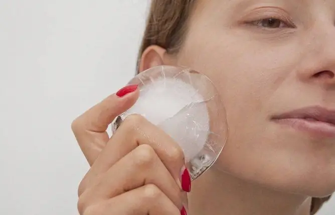
At home, the treatment of hematoma on the head after a bruise can be carried out only in mild cases. Therapy consists in applying cold to the injury site in the first hours after the injury. In the future, it is necessary to apply external agents to the area of impact, helping to dissolve the resulting bruise.
It is also important to report the injury to relatives, as the bleeding may be accompanied by further memory loss.
The adequacy of the therapy is assessed by the stabilization of the general condition and regression of the external manifestations of trauma.
Forecast
With an epidural form of pathology, in the case of timely treatment, the prognosis is good. The prognosis of subdural hemorrhage varies widely and depends on the size of the affected area and the duration of the injury.
After any significant blow to the head with loss of consciousness, or if any signs appear that may indicate a hematoma, see a doctor.
Video
We offer for viewing a video on the topic of the article.

Anna Kozlova Medical journalist About the author
Education: Rostov State Medical University, specialty "General Medicine".
Found a mistake in the text? Select it and press Ctrl + Enter.


