- Author Rachel Wainwright [email protected].
- Public 2024-01-15 19:51.
- Last modified 2025-11-02 20:14.
Malignant moles
The content of the article:
- Features
- Conditionally malignant neoplasms (precancer)
-
Cancer moles
- Melanoma
- Basal cell skin cancer
- Squamous cell carcinoma
- Video
Malignant moles are a group of skin cancers of epithelial origin that in most cases have an unfavorable outcome.
WHO proposed the following classification of malignant tumors of the skin and its appendages:
- epithelial tumors (skin cancer, adenocarcinoma of the sweat and sebaceous glands);
- connective tissue tumors (fibrosarcoma of the skin, dermatofibrosarcoma, leiomyosarcoma);
- vascular tumors (hemangioendothelioma);
- melanoma of the skin.

Malignant moles have a number of differences, however, only a doctor can determine exactly whether a mole is a cancerous tumor.
Features
They occur in people of any age and gender (some types have their own characteristics).
- People with pale skin are more prone to it.
- Direct relationship with the surrounding external environment. The provoking factors can be chemical carcinogens, physical (ultraviolet) or biological (human papillomavirus) effects.
- This type of neoplasm can be either primary (formed on intact skin) or secondary (occurs at the site of benign formations).
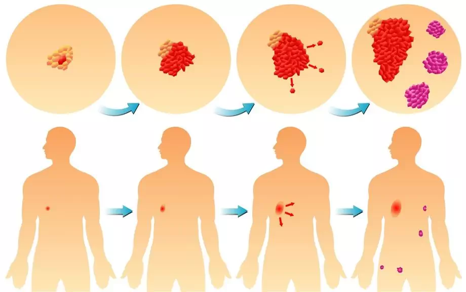
- Ways of metastasis in such cases to regional lymph nodes (distant metastases are rare and advanced cases).
- Skin cancer often occurs in open areas of the skin (about 60% of all cases). The trunk skin accounts for about 7-10%.
- For a long time they do not have any symptoms other than external manifestations (as the progression occurs, paraneoplastic syndrome occurs).
- Often occurs as a hereditary pathology (a history of cases of illness in close relatives).
These criteria are universal for any education in oncology. According to ICD 10, malignant neoplasms of the skin are coded with the code C 43-C 44.
How to determine a malignant mole or not
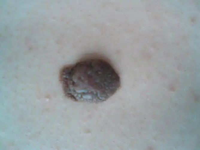
There are a number of important external signs of malignant moles (ABCDE method):
| Designation | Decoding |
| A - asymmetry | You can check this sign by drawing a conditional line through the supposed middle of the mole (in the case of malignant tumors, you will get two halves of different sizes). |
| B - edge (border) | In benign formations, the edges are smooth and clear, but with malignancy, the edges become torn like tongues of flame and lose their clarity. |
| С - color | The color of an ordinary mole should be uniform throughout. In the case of polychrome color and the predominance of a black shade, you must immediately contact a specialist. |
| D - diameter | The diameter of the malignant lesion, as a rule, exceeds 6 mm. |
| E - evolution of development | All benign formations change slightly over time (slightly enlarged, stretched). In the case of malignant forms, intensive growth is noted. |
Indirect poor diagnostic features:
- itching and redness;
- the disappearance of the skin pattern at the site of the lesion;
- peeling and swelling of the focus;
- hair loss;
- ulceration and change in density;
- bleeding and weeping surface;
- screenings from the main focus (satellites).
Conditionally malignant neoplasms (precancer)
A number of formations on the skin are conditionally malignant, since they have a high risk of transformation into cancer, even with minor exposure. These include:
- Bowen's disease. It is represented by a plaque with irregular edges and is localized on the skin of the face and trunk. The color ranges from reddish to yellow-brown. Size up to 10 cm. The focus is dense on palpation, covered with scales.
- Pigmented xeroderma. Hereditary predisposition is expressed. Has a high sensitivity to sunlight. It is represented by atrophied reddish patches of skin that resemble freckles. The size is variable. Often turns into multiple primary cancer.
- Paget's disease. It is localized in the area of the areola of the mammary glands and is represented by a reddish or pink spot. The surface of the formation may peel off. The diameter is variable.
- Keratoacanthoma (horny mollusc). It is localized in open areas of the skin. Has a hemispherical shape and dense consistency. It protrudes above the skin surface. The tumor is grayish-brown in color, and the stratum corneum above it is gray. In the center, as the disease progresses, a crater often appears.
- Senile keratosis. It occurs on any part of the body. Multiple patches of pale or brown color are characteristic. Local capillary hemorrhages (telangiectasias) are visible around the formations.
- Cutaneous horn. Formation of a dirty gray or brownish gray color, significantly protruding above the surface of the skin. Sizes up to 1 cm. Localization mainly on the skin of the face.
A special group that has a high risk of malignancy, i.e. malignancy, are melanoma-prone pigmented nevi:
- Dysplastic nevus. It is represented by a birthmark, which is located at the same level as intact skin. The shade ranges from brown to black. The edges are fuzzy.
- Border pigmented nevus. It is represented by a dark knot. Has a higher density than the surrounding tissue.
- Blue (blue) nevus. The knot is blue, bluish, dark brown. The edges are straight and crisp. Softly elastic on palpation, may not protrude above the skin surface.
- Nevus Oto. Combined damage to the skin, eyes (sclera, iris) and mucous membranes. It is localized in the zone of innervation of I and II branches of the trigeminal nerve (face, neck), in the photo it looks like a dirty gray spot of irregular outlines.
- Giant pigmented nevus. The formation exceeds 20 cm in diameter. The color is brown or grayish. Sometimes covered with hair.
- Melanosis Dubreya. The main element is a grayish-brown plaque with irregular outlines, dense on palpation.
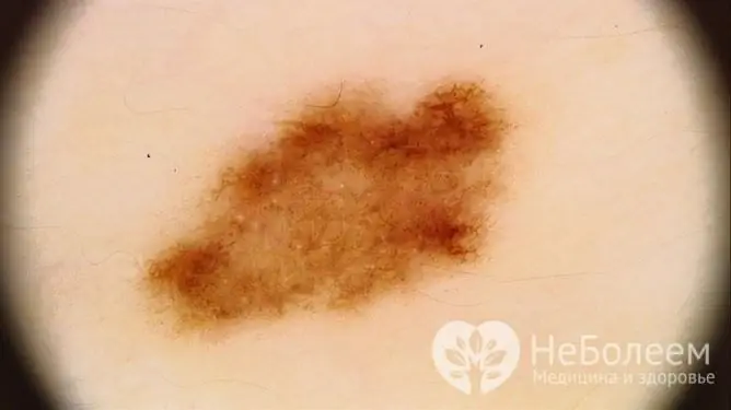
Dysplastic nevus refers to precancerous skin conditions
Due to the high degree of danger, those suspicious of malignant formation in most cases are subject to surgical treatment (removal) followed by biopsy, which makes it possible to understand the exact cellular composition of the tumor-like formation.
Cancer moles
Directly cancerous, that is, malignant moles are melanoma, basal cell and squamous cell carcinoma.
Melanoma
Another name is melanoblastoma. It is subdivided into several types.
Surface spreading (up to 70%)
- size up to 2 cm;
- brown and dark brown color is not uniform;
- localization on the skin of the chest, back and extremities;
- vertical and / or horizontal growth;
- level with intact skin;
- edges are uneven, wavy;
- various shapes (round, hexagonal);
- the surface is bumpy.
For a long time it is in a dormant state and often occurs against the background of pigmented nevi. At the initial stage of the disease, it is difficult to understand the specific nature of the pathological focus, which complicates timely diagnosis.
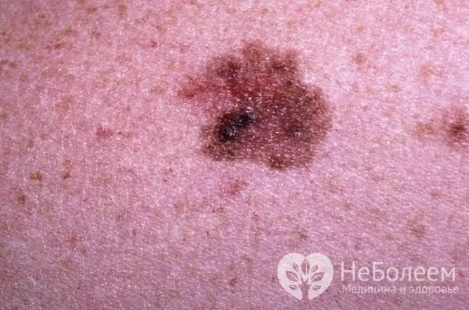
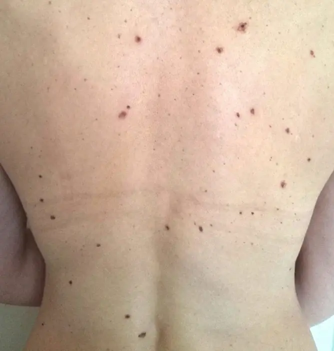
Melanoma may not differ from a normal birthmark, so you need to pay attention to any new birthmarks
Nodular (nodular)
It is considered the final stage of development of the surface form.
- localization - head and neck;
- polyp-type node;
- the surface is smooth, shiny, uneven;
- tendency to ulceration;
- colors similar to the surface form.
Lentigo-melanoma (melanotic freckles)
It always occurs de novo, that is, on unchanged skin. It can rarely occur against the background of nevi or other precancerous diseases. It appears more often in men in old age (over 70 years), in young people it is recorded in less than 1% of cases.
- localization - face and neck;
- multiple nodules;
- dark blue, dark brown and light brown;
- diameter 1.5-4 mm;
- edges are uneven, wavy.
Acral-lentiginous
- localization - palms, soles, nails;
- uneven edges;
- color gray and black.
More often occurs in persons of the Negroid race. It grows extremely slowly. Has a relatively favorable prognosis.
Basal cell skin cancer
Also subdivided into several forms.
Nodular
The most common type of pathology. Characteristics:
- dense multiple nodules prone to fusion;
- diameter 2-5 mm;
- localization - facial skin;
- color from pale pink to yellow-brown;
- has a tendency to ulceration.
Surface
Features:
- rounded superficial focus;
- diameter 1 cm or more;
- on the surface there are papillomatous growths, ulceration;
- occurs on the skin of the trunk and limbs;
- the color is yellowish.
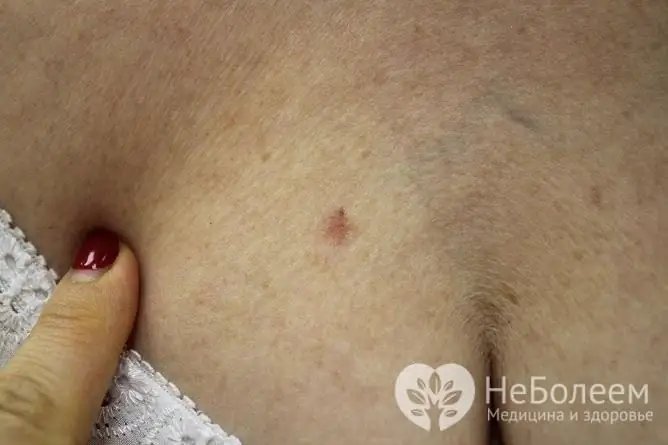
Basal cell skin cancer in the initial stages can take the form of a mole
Scleroderma-like
Relatively favorable shape.
Features:
- fast growth;
- does not protrude above normal skin at the beginning, and then, due to endophytic growth, it is pressed into the depths like a scar.
Fibroepithelial
Has a relatively favorable outcome.
Characteristics:
- knot shape (color varies widely);
- diameter 1-3 cm;
- tightly elastic;
- localized on the trunk.
Squamous cell carcinoma
Has three forms.
Superficial
Difficult to identify and the most common form.
Characteristics:
- single spot or nodule;
- the color is variable, often whitish;
- multiple crusts on the outbreak;
- protrudes above the surface of the skin;
- has a tendency to erosion.
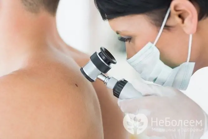
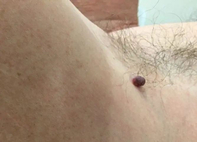
If you suspect a possible skin cancer, you should consult a dermatologist
Infiltrative (endophytic)
- multiple dense knots;
- crusted;
- a tendency to form deep ulcers.
Papillary form (exophytic)
- outwardly looks like cauliflower;
- rises above the skin;
- tendency to bleeding and ulceration.
Melanoma and all forms of squamous cell carcinoma most often develop regional and distant metastases in a short period of time (2-4 months), so they are most dangerous. Malignant moles do not make themselves felt for a long time, and for this reason the patient is often hospitalized in a hospital already against the background of pronounced paraneoplastic syndrome.
Video
We offer for viewing a video on the topic of the article.

Anna Kozlova Medical journalist About the author
Education: Rostov State Medical University, specialty "General Medicine".
Found a mistake in the text? Select it and press Ctrl + Enter.


