- Author Rachel Wainwright wainwright@abchealthonline.com.
- Public 2024-01-15 19:51.
- Last modified 2025-11-02 20:14.
Why do hanging moles appear and how to remove them
The content of the article:
- Features of hanging moles
- Localization features
- What causes hanging moles
- Why are hanging moles dangerous?
- Diagnostics
- Removing hanging moles at home
- Removal of hanging moles by a doctor
- Video
On the body of any person there are always various pigmented formations (moles, nevi). The reasons for their appearance, external manifestations and localization can vary significantly. The most unpleasant form from an aesthetic point of view among all epidermal neoplasms is hanging moles. In this article we will talk about why such benign growths appear on the surface of the skin and how you can get rid of them.

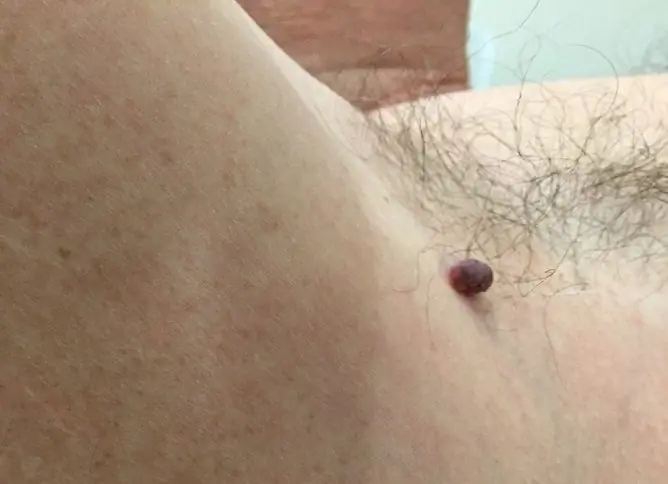
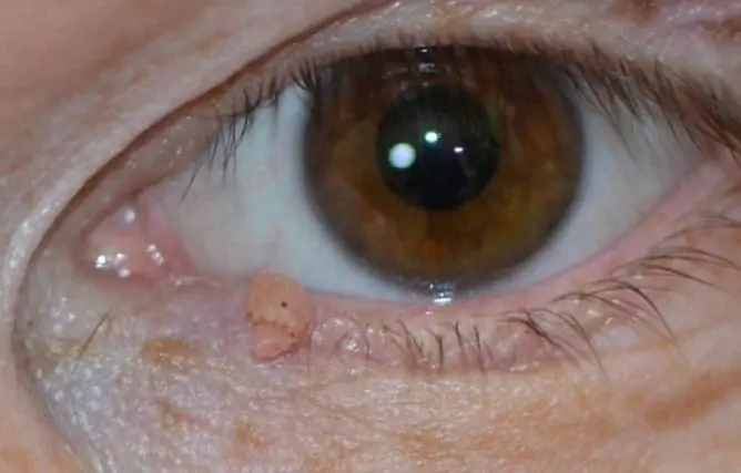
Hanging moles look unaesthetic, and when located in some anatomical zones, they are easily injured
Features of hanging moles
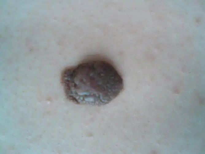
Hanging moles (nevi) mean epidermal neoplasms with a relatively small base, slightly protruding above the surface of the skin (varieties with a long stem are not considered to be considered). In some cases, their surface may consist of several slices and look like a cauliflower inflorescence.
Hanging nevi more often occur in men and women over the age of 30-35 and are acquired. Their color characteristics lie within a fairly wide range: from light beige, almost white, to dark brown.
The appearance of a convex pigmented formation does not always indicate the presence of any pathological process in the body, since many people, in the presence of such neoplasms, feel quite satisfactory, do not feel the slightest discomfort and do not present any complaints. But at the same time, this kind of benign tumors can create a number of problems for a person:
| Problem | Description |
| Aesthetic unattractiveness | The appearance of hanging moles is rather unattractive, therefore, when they are located in open areas of the body, for example, on the neck, they are considered by many patients as a serious cosmetic defect. |
| Risk of malignancy |
In the presence of any benign neoplasm, especially easily traumatized, there is a risk of its transformation into a malignant tumor. Signs of possible malignancy are: · Rapid growth of education; · Change of its color and / or shape; · The appearance of itching, burning. |
| Physical discomfort | The growths protruding above the surface of the skin are often rubbed with clothing and various accessories. This leads to their trauma, which is accompanied by the appearance of pain, itching, as well as an increased risk of transformation of the formation into malignant melanoma. In addition, damage to the integrity of the epithelium of the tumor surface can lead to the occurrence of purulent-infectious inflammation. |
Localization features
Unlike other types of nevi, which can be located anywhere on the human body, hanging moles have their favorite locations. Most often, they begin to grow in the following areas of the body:
| Epidermal zone | Features of the neoplasm |
| Neck | Education brings significant cosmetic discomfort to the patient. In addition, it is often traumatized by chains and collars of clothing. It is almost constantly exposed to ultraviolet radiation from sunlight. All these factors provoke tumor growth and increase the risk of malignancy. |
| Armpits |
The danger of this localization lies in the possible damage to the formation during shaving or epilation in this anatomical zone. Such injuries are especially dangerous for people with excessive sweating, since in this case the likelihood of microbial infection of the wound surface increases. |
| Back | Hanging moles are most common in the shoulder area. This makes it uncomfortable for women to wear shoulder bags and bras. |
| Groin | The most common type of localization. The lesions are prone to high trauma during sexual intercourse and when shaving the bikini area. |
What causes hanging moles
The following factors are the main causes of hanging pigmented lesions:
- Changes in hormonal levels. With any hormonal imbalance (puberty, pregnancy, hormonal therapy, menopause), the risk of the formation of benign pigmented skin structures increases. Therefore, they often form in women during pregnancy and are often located on the breast skin.
- Excessive insolation. Melanin is highly sensitive to ultraviolet rays, so their prolonged exposure to the skin (frequent sunbathing, visits to a solarium) can provoke the formation of various types of pigmented neoplasms.
- Human papillomavirus (HPV) infection. The virus accumulates in the cells of the epithelium and provokes their excessive division, which causes the formation of various outgrowths on the surface of the skin. In terms of histological structure, they differ from pigmented nevi, but many specialists do not share these structures, since they have similar clinical manifestations, prognosis, and also require the same treatment.
Why are hanging moles dangerous?
The main danger of any pigmented neoplasm is the possibility of its degeneration into a malignant skin tumor - melanoma. You can suspect such a transformation when the following signs appear:
- burning, pain or itching in the nevus area;
- rapid change in the shape and / or size of the formation;
- change in the consistency of the build-up, which is determined by palpation;
- discoloration of the surface of the nevus;
- the appearance on the surface of the neoplasm of ulcerated or bleeding areas.
If any of the listed symptoms appear, the patient should immediately consult a doctor - an oncologist or dermatologist. The specialist will conduct the studies necessary in each specific case, which will determine the level of oncological danger of the pigmented formation, and will recommend how to remove it.
Diagnostics
Currently, the diagnosis of pigmented formations is based on the data of the following methods:
- Cytology. If there are ulcerations or bleeding areas on the surface of the formation, then the doctor can make a smear-imprint and examine the cellular composition under a microscope.
- Histology. It is performed only in the conditions of an oncological dispensary. Under local anesthesia, complete excision of the formation is performed with the obligatory capture of the surrounding healthy tissues. The wound is sutured, and the resulting tissue is sent for histological analysis.
- Epiluminescence microscopy. With the help of a special device - a dermatoscope, intravital microscopy of a skin neoplasm is performed.
- Computer diagnostics. A series of sighting photos of a pigmented nevus is performed with a high-resolution camera. The resulting images are processed using a computer program.
Removing hanging moles at home
Some people, when hanging moles appear, prefer not to see a doctor and get rid of them using traditional medicine methods. It is categorically not recommended to do this, since such attempts are always associated with a high risk of developing various complications. Let's consider some of these methods:
- Tying the base of the convex formation with silk thread. The method is based on the fact that after ligation of the leg, the blood supply to the formation stops. Over time, it mummifies (dries up) and disappears. However, with such attempts at treatment, there is a high risk of infection with the development of purulent complications, the formation of rough scars.
- Moxibustion of the formation with celandine juice, vinegar essence. Attempts to remove skin growths in such ways are often accompanied by deep burns of the surrounding soft tissues.
- Ingestion of decoctions of poisonous plants (hemlock, celandine). These plants contain a huge amount of various biologically active substances. Ingestion of phytopreparations based on them can cause serious poisoning, which can even lead to death.
Patients should be well aware of the potential consequences of self-medication. In addition to all of the above, improper removal of pigmented neoplasms can cause their malignancy.
Removal of hanging moles by a doctor
If you need to get rid of a pigmented nevus, it is best to see a doctor.

To remove a mole, you should contact a specialist
Currently, to eliminate these types of skin formations, specialists practice the following methods:
- surgical excision with the imposition of skin sutures - the operation is indicated for patients with melanoma-prone neoplasms;
- electrocoagulation - the advantages of this method are the ability to perform a tissue biopsy for subsequent histological analysis, as well as the bloodlessness of the procedure and rapid wound healing;
- cryodestruction - destruction of a tumor by deep freezing it with liquid nitrogen (rough scars may remain);
- laser radiation is one of the most effective, safe and low-traumatic ways to remove hanging moles, after which no visible scars remain on the skin;
- radio wave surgery - the method is based on the use of high frequency radio waves, minimally injuring the surrounding soft tissues.
The procedure for removing pigmented nevi is performed under local anesthesia, so it does not cause discomfort or pain in patients.
Video
We offer for viewing a video on the topic of the article.

Elena Minkina Doctor anesthesiologist-resuscitator About the author
Education: graduated from the Tashkent State Medical Institute, specializing in general medicine in 1991. Repeatedly passed refresher courses.
Work experience: anesthesiologist-resuscitator of the city maternity complex, resuscitator of the hemodialysis department.
Found a mistake in the text? Select it and press Ctrl + Enter.

