- Author Rachel Wainwright wainwright@abchealthonline.com.
- Public 2023-12-15 07:39.
- Last modified 2025-11-02 20:14.
Tumor markers: decoding

Window markers are specific protein substances produced in excess by cancer cells, found in the blood or urine of patients with cancer or those at risk. Tumor growth markers are complex protein molecules with a carbohydrate or peptide component. Basically, tumor markers are proteins, enzymes, hormones, protein breakdown products of a malignant neoplasm, as well as metabolic products and antigens associated with neoplasm. Tumor markers differ from compounds that are normally produced by healthy cells qualitatively (tumor-specific markers) and quantitatively (associated with neoplasms, but present in healthy cells). Tumor markers are formed on the surface of modified tumor cells. Some of them are secreted into the bloodstream,thanks to which tumor markers become available for identification by blood sampling. Currently, there are about 200 types of tumor growth markers, but only 20 of them have diagnostic value. When determining tumor markers, decoding will allow to establish the localization, type of neoplasm, stage of the tumor process.
Tumor markers by their nature are divided into the following classes:
- Hormones - ACTH, human chorionic gonadotropin;
- Immunological - antigens and antibodies associated with neoplasm;
- Protein decay products of neoplasms;
- Metabolic products - free DNA, creatine, polyamines;
- Plasma proteins - beta-2-microglobulin, ferritin, ceruloplasmin;
- Enzymes - lactate dehydrogenase, phosphatase.
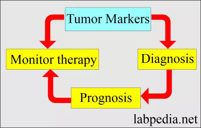
Identification of tumor markers, deciphering their content in the blood helps to solve the following tasks:
- Differential diagnosis of neoplasms of any nature at any stage of development;
- Determination of the effectiveness of the applied treatment, as well as possible changes in therapy;
- Monitoring the course of the disease;
- Early detection of relapses and metastases (6 months before the first clinical manifestations).
At the same time, the dynamics of the growth of the tumor marker, the decoding of the change in the level of the substance content is of greater importance than a single indicator. Currently, no tumor marker has been identified that has 100% specificity and is associated with a specific organ. However, when tumor markers are detected, decoding will allow, with a high degree of probability, to determine which organ is affected and what the extent of the lesion is.
Decoding the analysis for tumor markers: types of tumor markers, the rate of the content of tumor markers in the blood
The material for the analysis for tumor markers is the patient's venous blood or urine. An analysis for tumor markers is prescribed if a neoplasm is suspected. Upon receipt of the results, only the attending physician should decipher the analyzes for tumor markers, since the presence of tumor markers in the material is not sufficient for making a diagnosis. There is a concept of the norm for the content of tumor markers, the decoding of which should be carried out only by an oncologist.
When conducting analyzes for tumor markers, decoding should be carried out in accordance with the following rules:
- When identifying a tumor marker, decoding first of all determines a deviation from the norm, on the basis of which a decision is made to conduct a repeated analysis;
- The diagnosis in the presence of tumor markers in the patient's blood is made on the basis of the clinical picture, and not on the basis of a separate indication of the level of the tumor marker.
The most commonly identified tumor markers are:
- CA 125 is a marker of ovarian cancer. The norm is 0 - 30 IU / ml;
- CEA - cancer embryonic antigen, which is a tumor marker for rectal cancer. The normal value ranges from 0 - 5ng / ml;
- AFP (alpha-fetoprotein) is a marker of hepatocellular liver cancer. The norm is 0 - 5 IU / ml;
- PSA is a tumor marker for prostate cancer (prostate-specific antigen). The content of this tumor marker in the range of 0-4ng / ml is normal.
When tumor markers are detected, the decoding can indicate not only the presence of malignant tumors, but also the usual inflammatory processes, diseases, dysfunction of organs or their systems.
Tumor marker CA 125: decoding, possible diseases
The tumor marker CA 125 is a glycoprotein with a molecular weight of 220 kD. This substance is present in the epithelial tissues of the ovaries, stomach, intestines, pancreas, kidneys, gallbladder, bronchi. The tumor marker CA 125 can also be detected in the blood plasma of pregnant women and in breast milk. The CA 125 tumor marker is used as a secondary marker in the diagnosis of pancreatic cancer. An increased concentration of the CA 125 tumor marker, the decoding of which makes it possible to determine the presence of ovarian neoplasms, may also indicate the presence of inflammatory processes in the appendages. The basis for the analysis for the CA 125 tumor marker are:
- diagnostics of ovarian pathology, screening of neoplasms;
- diagnosed adenocarcinoma of the pancreas.
In the presence of the CA 125 tumor marker, the decoding indicates either oncopathology or somatic pathology. With an increased level of this tumor marker, decoding determines oncological pathological processes:
- Cancers of the ovaries (up to 80% of cases), fallopian tubes, uterus, endometrium;
- Liver cancer;
- Lung cancers;
- Cancers of the stomach, rectum, pancreas;
- Malignant formations of the breast;

An increased concentration of the CA 125 tumor marker may also indicate somatic pathology:
- Endometriosis;
- Inflammatory processes in the uterus and appendages;
- Ovarian cystic formations;
- Pleurisy, peritonitis;
- Liver cirrhosis, chronic forms of hepatitis;
- Chronic pancreatitis;
- Autoimmune pathology.
When identifying this tumor marker, decoding should not take into account only the level of the substance content. The increased concentration of CA 125 requires additional tests for tumor markers, the decoding of which together will make it possible to make an accurate diagnosis. When identifying window markers, the decoding carried out independently is not informative, since it is impossible to determine what level of the tumor marker content corresponds to a particular disease.
CEA tumor marker: decoding, possible diseases
CEA tumor marker is a glycoprotein belonging to the group of carcinofetal antigens that are formed during embryonic development. An increased concentration of CEA indicates possible malignant neoplasms.
When determining an increased concentration of the CEA tumor marker, the decoding may indicate cancer of the ovaries, breast, lungs, rectum, stomach, pancreas. Due to the fact that in the presence of the CEA tumor marker, it is impossible to decipher and diagnose only by the content of this tumor marker, the patient is prescribed a number of tests based on the general clinical picture.
YouTube video related to the article:
Found a mistake in the text? Select it and press Ctrl + Enter.


