- Author Rachel Wainwright [email protected].
- Public 2024-01-15 19:51.
- Last modified 2025-11-02 20:14.
Blood test for tumor markers
The content of the article:
- Donating blood for analysis for tumor markers
- Norms of blood test indicators for tumor markers
-
What does the blood test for tumor markers say and what does it show?
- Alpha-fetoprotein
- Cancer-embryonic antigen
- Ovarian tumor marker CA-125
- Breast tumor marker CA 15-3
- Pancreatic tumor marker CA 19-9
- Prostate-specific antigen
- Human chorionic gonadotropin
A blood test for tumor markers is prescribed if a tumor is suspected. Those who are at risk of developing malignant tumors are recommended to undergo the study annually. The risk group includes people with a genetic predisposition to cancer, chronic diseases, precancerous pathologies, as well as living in ecologically unfavorable regions or working in hazardous industries. In the presence of oncological disease, the analysis is carried out for monitoring purposes.

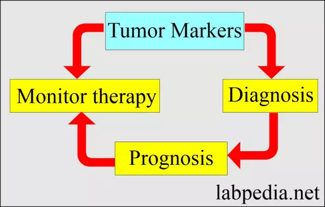
A blood test for tumor markers is used for early diagnosis of certain types of tumors, control of treatment, detection of metastases and relapse A blood test for tumor markers is used for early diagnosis of certain types of tumors, control of treatment, detection of metastases and relapse
Tumor markers are metabolic products of tumor formation, as well as substances produced by normal tissues of the body in response to the invasion of cancer cells. In the body of healthy people, some tumor markers are present in small quantities; an increase in their concentration in the blood and urine of patients indicates the development of cancer with a high probability. In some cases, tumor markers increase in some non-cancer diseases.
To prescribe an analysis and interpret the results of the study, you must contact a qualified specialist who will explain what the blood test for tumor markers is talking about and what the blood test shows, how the material is taken and how the analysis is done, as well as how to prepare for it.
Donating blood for analysis for tumor markers
Blood sampling for analysis is carried out in the morning on an empty stomach, after the last meal should pass 8-12 hours. Whether it is possible to take a blood test for tumor markers at other times of the day should be clarified in a specific laboratory and with the doctor who ordered the study. For analysis, blood is taken from a vein.
For a blood test for tumor markers, preliminary preparation is required. A few days before blood sampling, fatty, fried and spicy foods, alcoholic beverages should be excluded from the diet. Before donating blood, you should not smoke during the day; emotional and physical stress should be eliminated within 30 minutes. In case of taking medications, you need to consult a doctor and find out if there is a need to cancel them. It is also advisable to agree with the doctor on which days it is better to take the test to obtain the most reliable research result (for example, in women, the results of some tests depend on the phase of the menstrual cycle).
A prostate-specific antigen (PSA) test is possible no earlier than 1-2 weeks after digital rectal examination or prostate massage, transrectal ultrasound and other hardware diagnostic methods. How long you need to wait after each specific manipulation should be checked with your doctor. In addition, two days before the study, it is necessary to exclude sexual contact and serious physical activity.
Norms of blood test indicators for tumor markers
The table shows the norms of the most frequently determined tumor markers. In different laboratories, depending on the test method and the adopted units of measurement, normal values and may differ.
Norms of blood test indicators for tumor markers
Index Reference values Alpha-fetoprotein (AFP) Men and non-pregnant women - up to 2.64 IU / ml
pregnant women - 23.8-62.9 IU / ml (depending on the duration of pregnancy)
Cancer-embryonic antigen (CEA) Men - up to 3.3 ng / ml non-smokers, up to 6.3 ng / ml smokers
women - up to 2.5 ng / ml non-smokers, up to 4.8 ng / ml smokers
Ovarian tumor marker CA-125 Up to 35 U / ml Breast tumor marker CA 15-3 Up to 32 U / ml Pancreatic tumor marker CA 19-9 Up to 37 U / ml Prostate-specific antigen common Up to 4 ng / ml Human chorionic gonadotropin (hCG) total beta subunit Men - up to 2.5 U / l
Women - up to 5 U / l
What does the blood test for tumor markers say and what does it show?
Alpha-fetoprotein
Alpha-fetoprotein (AFP, AFP) is an embryonic serum protein that is produced during the development of the embryo and fetus. Alpha-fetoprotein is structurally similar to serum albumin in adults. Its function is to prevent rejection of the fetus by the mother's body. In children, the AFP level in the blood is high at birth, then progressively decreases and reaches normal adult values by the age of two. High alpha protein levels in adults are a sign of pathology.
Alpha-fetoprotein is one of the main indicators of chromosomal abnormalities and fetal abnormalities during intrauterine development. Its determination in pregnant women is often prescribed in combination with ultrasound examination, determination of the level of human chorionic gonadotropin and free estriol, which makes it possible to assess the risks of developing pathologies in the fetus in combination.
An increase in the level of alpha-fetoprotein in a pregnant woman may indicate multiple pregnancy, necrosis of the fetal liver against a background of a viral infection, open defects in the development of the neural tube, umbilical hernia, Meckel-Gruber syndrome.
In men and non-pregnant women, indications for prescribing an analysis for alpha-fetoprotein are usually the detection of metastasis, assessment of the effectiveness of therapy for malignant neoplasms, and determination of the risk of developing oncopathology (in persons with chronic viral hepatitis, liver cirrhosis, etc.).
An increase in the concentration of alpha-fetoprotein in men and non-pregnant women occurs with hepatocellular carcinoma, liver metastases of tumors of other localizations, neoplasms of the testicles, lungs, stomach, pancreas, and large intestine. AFP slightly increases in chronic hepatitis, cirrhosis, alcoholic liver damage.
A decrease in the level of alpha-fetoprotein after a course of treatment or removal of a neoplasm means an improvement in the patient's condition. A decrease in AFP in the blood of a pregnant woman may indicate the presence of chromosomal abnormalities in the fetus (Edwards or Down syndrome), an incorrectly defined gestational age (overestimated), cystic drift, spontaneous abortion, and fetal death.
Cancer-embryonic antigen
Cancer-embryonic antigen (CEA, CEA, carcinoembryonic antigen) is an embryonic glycoprotein that is produced in the tissues of the digestive tract of the embryo and fetus. Its function is to stimulate cell proliferation. After the birth of a child, the synthesis of the cancer-embryonic antigen is suppressed; it is present in a small amount in the blood of an adult. An increase in CEA occurs during the development of a tumor in the body and reflects the progression of the pathological process.
A blood test for cancer-embryonic antigen is indicated in the diagnosis of medullary carcinoma, cancer of the pancreas, stomach, colon and rectum, in the assessment of the ongoing cancer treatment, and is also used for the early detection of malignant tumors during screening of risk groups.
An increase in CEA concentration does not necessarily indicate cancer; it occurs in intestinal polyposis, Crohn's disease, ulcerative colitis, hepatitis, cirrhosis, liver hemangioma, pancreatitis, cystic fibrosis, pneumonia, pulmonary emphysema, tuberculosis, renal failure. With these pathologies, the level of tumor marker usually does not exceed 10 ng / ml.
In addition, the concentration of CEA increases in cancer of the lung, breast, pancreas, ovaries, prostate, liver, thyroid gland, colorectal carcinoma, metastases to the liver or bone tissue.
An increase in the level of cancer-embryonic antigen after a decrease in its concentration may indicate relapses and tumor metastasis. The concentration of the cancer-embryonic antigen in the blood is influenced by smoking and drinking.
Ovarian tumor marker CA-125
CA-125 is a glycoprotein that is used as a marker of non-mucinous epithelial forms of ovarian malignant tumors and their metastases. In the case of heart failure, the level of CA-125 correlates with the concentration of natriuretic hormone, which can serve as an additional criterion for determining the severity of the patient's condition.
A blood test for the CA-125 tumor marker is prescribed during the diagnosis of ovarian cancer and its recurrence, pancreatic adenocarcinoma, as well as to assess the quality of treatment and prognosis.
The level of CA-125 increases in malignant neoplasms of the ovaries (in about 80% of patients, but at the initial stage - only in 50%), uterus, fallopian tubes, breast, rectum, stomach, pancreas, liver, lungs. An increase in CA-125 can also occur with inflammation in the small pelvis or abdominal cavity, autoimmune diseases, viral hepatitis, liver cirrhosis, ovarian cyst, during menstruation. A slight increase in the tumor marker can be observed in the first trimester of pregnancy in the absence of any pathology.

Tumor marker CA-125 is used in the diagnosis of ovarian cancer Tumor marker CA-125 is used in the diagnosis of ovarian cancer
Breast tumor marker CA 15-3
CA 15-3 is a glycoprotein produced by breast cells. In the early stages of breast tumors, the tumor marker exceeds normal values in about 10% of cases; in the presence of metastases, an increase in CA 15-3 level is observed in 70% of patients. An increase in its concentration can outpace the onset of clinical symptoms by 6-9 months. For the diagnosis of breast cancer at the initial stage, the 15-3 tumor marker is not sufficiently sensitive, but with already detected cancer, it makes it possible to monitor the course of the disease and evaluate the effectiveness of the treatment. The diagnostic value of the CA 15-3 tumor marker increases when it is determined in combination with a cancer-embryonic antigen.
Oncomarker CA 15-3 allows for the differential diagnosis of malignant neoplasms of the breast and benign mastopathy.
The concentration of the CA 15-3 tumor marker increases in malignant neoplasms of the breast, rectum, liver, stomach, pancreas, ovaries and uterus, as well as in cirrhosis, viral hepatitis, rheumatic and autoimmune diseases, pathologies of the lungs and kidneys. In addition, a slight increase in CA 15-3 levels occurs during pregnancy.
Pancreatic tumor marker CA 19-9
CA 19-9 is a sialoglycoprotein that is produced in the gastrointestinal tract, salivary glands, bronchi, lungs, prostate gland, but is used primarily for the diagnosis of pancreatic cancer.
A blood test for the CA 19-9 tumor marker is usually prescribed when a malignant process in the pancreas is suspected, to assess the effectiveness of its treatment and determine the risk of recurrence. Sometimes CA 19-9 is used for suspected malignant tumors of other localization.
An increase in the level of CA 19-9 occurs in cancer of the pancreas, gallbladder, liver, stomach, breast, ovaries, uterus, as well as colorectal cancer. A slight increase in the tumor marker may indicate cholecystitis, hepatitis, gallstone disease, cirrhosis of the liver, autoimmune diseases, and in addition, it occurs in about 0.5% of clinically healthy people.
Prostate-specific antigen
Prostate-specific antigen (PSA, PSA) is a protein produced by prostate cells that serves as a marker for prostate cancer. Total PSA is the sum of the free and protein-bound fractions.
Indications for analysis for prostate-specific antigen are monitoring the course of prostate cancer, detecting metastasis and monitoring treatment, assessing the condition of patients with benign prostatic hypertrophy in order to detect possible malignancy early, prophylactic examination of men at risk (over the age of 50, with a genetic predisposition etc.).
The content of prostate-specific antigen in the blood increases in prostate cancer (in about 80% of patients), prostate adenoma, infectious and inflammatory processes, heart attack or prostate ischemia, trauma or surgery on the prostate gland, acute renal failure, acute urinary retention.
Physiological increase in the level of prostate-specific antigen occurs with constipation, after sexual intercourse, rectal digital examination of the prostate, as this often damages the capillaries of the prostate gland.
If the level of total PSA in the blood is high, the level of the free fraction should be determined to differentiate between benign and malignant processes.
Human chorionic gonadotropin
Human chorionic gonadotropin (hCG) is a hormone that begins to be produced by the chorionic tissue on the 6-8th day after the fertilization of the egg and is one of the most important indicators of the presence and normal course of pregnancy. The hormone consists of alpha (common for luteinizing, follicle-stimulating and thyroid-stimulating hormones) and beta (specific for hCG) subunits. Determination of the beta-subunit level allows you to diagnose pregnancy as early as a week after conception.
In non-pregnant women and men, the appearance of hCG in the blood indicates a neoplasm that produces a hormone. These can be tumors of the lungs, kidneys, testicles, organs of the gastrointestinal tract. An increase in the concentration of chorionic gonadotropin is noted with cystic drift, chorionic carcinoma.
YouTube video related to the article:

Anna Aksenova Anna Aksenova Medical journalist About the author
Education: 2004-2007 "First Kiev Medical College" specialty "Laboratory Diagnostics".
Found a mistake in the text? Select it and press Ctrl + Enter.


