- Author Rachel Wainwright wainwright@abchealthonline.com.
- Public 2023-12-15 07:39.
- Last modified 2025-11-02 20:14.
Decoding a clinical blood test
The content of the article:
- Clinical blood test: decoding the analysis
- Erythrocytes
- Erythrocyte indices
- Erythrocyte sedimentation rate (ESR)
- Hemoglobin
- Leukocytes
- Platelets
- Donating blood for analysis
CBC is a basic laboratory test used to diagnose and monitor the treatment of many diseases. It is usually prescribed during the initial examination by any specialist.

Blood for clinical analysis is usually taken from a finger, but in some cases from a vein
Clinical blood test: decoding the analysis
A clinical blood test can be abbreviated, containing indicators of the total number of leukocytes, hemoglobin and erythrocyte sedimentation rate, and expanded, which indicates the results of studying all blood elements and an expanded leukocyte formula. If necessary, the number of platelets and reticulocytes is determined.
Erythrocytes
Red blood cells are red blood cells containing hemoglobin. Red blood cells are formed in the bone marrow, from where they enter the bloodstream, and their lifespan is 120 days. They are replaced by young red blood cells called reticulocytes. The reticulocyte count is used to estimate the rate of renewal of the blood composition.

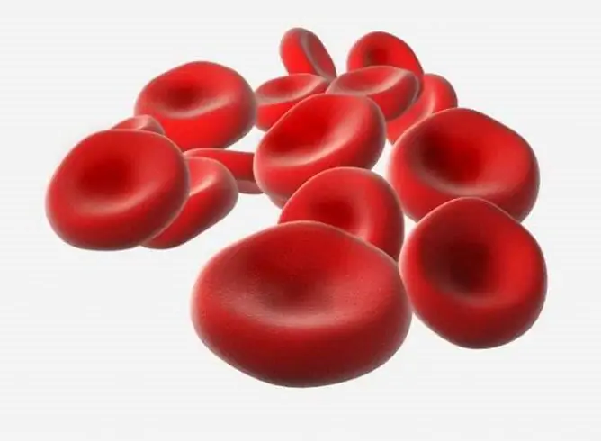
Red blood cells are red blood cells whose function is to carry oxygen to tissues
The main function of erythrocytes is to transport oxygen and carbon dioxide between the lungs and tissues of other organs. For the synthesis of red blood cells, a sufficient amount of vitamin B12, iron and folic acid is required.
An excess of red blood cells (erythrocytosis) is observed with dehydration of the body, which most often develops with indomitable vomiting, diarrhea, high temperature. The number of erythrocytes increases in diabetes, diseases of the heart and blood vessels, lungs, liver, kidneys, bone marrow dysfunction - erythremia, etc. Physiological erythrocytosis is observed in people under oxygen deficiency, stress, as well as in hyperhidrosis, increased physical activity.
The reason for a decrease in the level of red blood cells (erythropenia) can be anemias of various origins, renal diseases that increase the level of erythropoietin, blood loss, conditions accompanied by impaired formation or increased destruction of red blood cells, liver diseases associated with tissue changes, erythremia, hypothyroidism, oncological diseases, autoimmune pathologies.
In addition to the total number of erythrocytes, a number of their characteristics are examined in a clinical blood test: shape, size, quantity, hematocrit, hemoglobin content, and others.
Erythrocyte indices
The state of erythrocytes can be quantitatively characterized using erythrocyte indices:
- hematocrit is the percentage that red blood cells make up of the total blood volume;
- average erythrocyte volume (MCV) - the ratio of hematocrit to the number of erythrocytes;
- the average amount of hemoglobin in an erythrocyte (MCH) - this parameter is similar to the color indicator, but more accurately reflects the level of hemoglobin in an erythrocyte;
- distribution of erythrocytes in size (RDW) - characterizes fluctuations in cell volume within the population;
- the average concentration of hemoglobin in erythrocytes (MCHC) is a concentration index that determines the ratio of the amount of hemoglobin to the volume of the cell.
These calculated values, in combination with other indicators of the clinical blood test, are used for the differential diagnosis of anemia and a number of other diseases.
Erythrocyte sedimentation rate (ESR)
Erythrocytes are heavier than blood plasma, therefore, in a vertically installed test tube with an added anticoagulant, after a while, blood is divided into two layers: the upper one is transparent plasma and the lower one is erythrocytes that have settled under the influence of gravity. The erythrocyte sedimentation rate depends on many factors: the physicochemical properties of red blood cells, blood viscosity, the content of bile pigments and acids in it, acid-base balance, the balance of cholesterol and lecithin. In the inflammatory process, the erythrocyte sedimentation rate changes.

The erythrocyte sedimentation rate changes with the development of various diseases
What does increased ESR mean? It can be a sign of inflammation, infectious disease, myocardial infarction, kidney disease, trauma, cancer pathology. The physiological cause of an increase in ESR can be pregnancy, the postpartum period, menstruation, and undergone surgery. The increase in the indicator is also affected by the intake of estrogens, glucocorticoids.
A low erythrocyte sedimentation rate can be the result of overhydration, muscle dystrophy, unbalanced nutrition, and hormonal drugs.
Hemoglobin
Hemoglobin is a complex protein, a component of erythrocytes. It is he who gives the blood a rich scarlet color thanks to the iron atoms it contains. Hemoglobin is a respiratory blood pigment, its main function is to provide the body with oxygen. Hemoglobin transports oxygen and carbon dioxide between the lungs and body tissues, and maintains the pH of the blood.
The hemoglobin level decreases as a result of blood loss, a decrease in the saturation of blood with red blood cells in anemia. Malnutrition, disruption of the gastrointestinal tract can cause a decrease in hemoglobin. With a decrease in the amount of hemoglobin, oxygen starvation of cells begins, and acidosis develops.
An increased level of hemoglobin in the blood can be a sign of cardiovascular diseases with impaired vascular function, pathologies of the hematopoietic system with impaired erythropoiesis, blood clots, intestinal obstruction, neoplasms in the kidneys, liver, central nervous system, ovaries. An increase in hemoglobin in the blood can occur as a result of dehydration, burns, chemical poisoning, excessive physical exertion, stress. The level of glycated hemoglobin rises in diabetes mellitus and iron deficiency states.
Interpretation of the analysis results allows diagnosing diseases that are associated with a change in the amount of hemoglobin in erythrocytes, tracking metabolic processes in the body, and assessing the risks of complications of diabetes mellitus. The hemoglobin content in the blood of women is lower than that of men.
Leukocytes
Leukocytes are white blood cells that perform an immune function, involved in immune and inflammatory responses. Leukocytes are produced in the lymph nodes and red bone marrow.
An increase in the level of leukocytes accompanies inflammatory and infectious diseases, heart attacks of internal organs, intoxication, bleeding, tumor processes of hematopoietic tissue. Physiological causes of leukocytosis can be pregnancy, childbirth, menstruation, excessive physical activity, food intake, exposure to stress, cold, excessive sun exposure. An increase in the level of leukocytes in the blood can be affected by surgery, taking glucocorticoid drugs. In infants, leukocytosis is a normal stage in the formation and development of the immune system.

Leukocytes, or white blood cells, perform an immune function in the body, i.e. a protective function
A decrease in the number of leukocytes can be a sign of rheumatoid arthritis, lupus erythematosus, hypo- and aplasia of the bone marrow, leukopenic forms of leukemia. Leukopenia is often observed with anaphylactic shock, hypovitaminosis, general depletion of the body. A decrease in leukocytes can cause exposure to ionizing radiation, the use of antibacterial drugs, cytostatic, thyreostatic, antispasmodic, antiepileptic and non-steroidal anti-inflammatory drugs.
Leukocyte formula speaks about the relative (percentage) content of different types of leukocytes in the total blood volume. The study of the leukocyte formula gives an idea of the severity of the disease, the effectiveness of the treatment.
There are five types of leukocytes, each of which differs in its physical and functional characteristics:
- neutrophils are the most numerous type of leukocytes. provide the body's main defense against bacteria, fungi and protozoa. Localized in the foci of inflammation, neutrophils surround and phagocytose bacterial agents and tissue degradation products with the help of lysosomal enzymes. Depending on the stage of cell maturation, neutrophils are divided into subgroups. The ratio of subgroups to each other is called a neutrophilic formula, its shift to the left with an increase in the number of immature forms of neutrophils is a sign of an inflammatory process. Lack of neutrophils leads to chronic infections, weakening of the immune system. An increase in the number of neutrophils in the blood (neutrophilic leukocytosis) can be caused by the development of an acute infectious disease, a chronic inflammatory process, and some myeloproliferative diseases;
- lymphocytes - play a central role in immune responses, are responsible for acquired immunity, and promote tissue regeneration. Lymphocytes differentiate into subgroups with different functions: T cells, B cells, NK cells (natural killer cells);
- monocytes - have the highest ability to phagocytosis, absorb particles of foreign physical agents and foreign cells in the blood. A decrease in the level of monocytes may be due to the development of anemia, purulent lesions, leukemia. The decrease in monocytes can also be affected by surgical operations, taking steroid drugs;
- eosinophils - carry out extracellular destruction of parasitic organisms, they are able to phagocytose microbial cells, fight against particles that carry allergens in the focus of inflammation. When activated, eosinophils accumulate and release inflammatory mediators. Eosinophils are involved in the pathogenesis of bronchial asthma, other allergic diseases, helminthic and protozoal invasion. A decrease in the level of eosinophils means that inflammatory, purulent processes, or intoxication with salts of heavy metals develop in the body;
- basophils - secrete inflammatory mediators that increase vascular permeability, regulate blood coagulation and vascular permeability, play an important role in immediate allergic reactions. An increased number of basophils in the blood may indicate the development of myeloid leukemia, an allergic or infectious disease, endocrine pathology, hemolytic anemia, and malignant neoplasm. The reason for the increased values of basophils can be the condition after splenectomy, as well as the intake of hormonal drugs.
Platelets
Platelets are blood cells that support the functioning of blood vessels and are essential for normal hemostasis. Platelets are secreted in the bone marrow stem cell and are responsible for blood clotting and regeneration of damaged vessels. The number of platelets characterizes the body's ability to stop bleeding, this parameter is determined when assessing the blood coagulation system, when diagnosing thrombosis and malignant diseases of the bone marrow.

Platelets are responsible for blood clotting
An increased level of platelets is observed in inflammatory processes, tuberculosis, oncological diseases, lymphogranulomatosis, after undergoing surgical interventions. High platelet counts in the blood can be caused by certain medications. A decrease in the number of platelets occurs in acute leukemia, collagenosis, and cirrhosis of the liver.
Thrombocritis is an index that, as part of a clinical blood test, shows the percentage of platelet mass in the total blood volume.
Donating blood for analysis
Typically, blood for clinical analysis is taken from a finger, but in some cases, blood from a vein may be required. Blood sampling is recommended in the morning on an empty stomach, at least 8 hours should elapse since the last meal. Preparation for donating blood involves the elimination of psycho-emotional and physical stress, as well as drugs. The day before donating blood, you must exclude the use of alcohol, an hour - smoking.
YouTube video related to the article:

Anna Kozlova Medical journalist About the author
Education: Rostov State Medical University, specialty "General Medicine".
Found a mistake in the text? Select it and press Ctrl + Enter.


