- Author Rachel Wainwright wainwright@abchealthonline.com.
- Public 2023-12-15 07:39.
- Last modified 2025-11-02 20:14.
Dysplasia of the hip joint
The content of the article:
- Causes and risk factors
- Forms of the disease
- Symptoms
- Diagnostics
- Treatment
- Possible complications and consequences
- Forecast
Dysplasia of the hip joint (from ancient Greek δυσ - "violation" and πλάθω - "form") is a pathology caused by a violation of the formation of elements of the joint itself and its auxiliary apparatus in the prenatal period.

Stages of joint dysplasia
The hip joint is the largest and most heavily loaded movable joint in the body. Its articular surfaces are made by the acetabulum of the pelvic bone and the head of the femur, fixation of which (prevention of upward displacement) is provided by the acetabular lip (another name - "limbus") - a cartilaginous element that limits the cavity.
Anatomically and physiologically complete interposition of the articulating surfaces is provided by the articular capsule and ligamentous apparatus. The correct structure of the auxiliary structures protects the joint from subluxation and dislocation (displacement of the articular surfaces relative to each other) under conditions of increased stress.
During the neonatal period, the hip joint, even in healthy children, presents a rather unstable biomechanical structure, which is due to a number of age characteristics:
- flattened, shallow acetabulum;
- larger size of the femoral head in relation to the size of the cavity;
- poorly developed muscular frame in the gluteal region;
- insufficient compaction of the joint capsule.
Additional development of the joint occurs during the first year of life, almost completing by the age when the child begins to move independently.
With dysplasia of the anatomical structures that form the joint and its auxiliary apparatus, there is a high probability of abnormal development of the hip joint in the first months of life; as a result, the risk of trauma increases, the appearance of difficult-to-correct defects in gait, posture, and subsequent disability is possible.
The incidence of pathology in different countries is from 2 to 10%. Girls are more susceptible to the disease (8 out of 10 cases), the left hip joint is involved in the process most often - more than half of all identified dysplasias, pathologies of the right joint and combined (with damage to both joints) occur equally, in approximately 20% of patients. In the case of diagnosing a breech presentation of the fetus, the risk of dysplasia increases 10 times.
Causes and risk factors
The main reason for the pathological condition is connective tissue dysplasia, manifested by increased extensibility of connective tissue structures, a decrease in their strength.

Influenza, SARS, rubella transferred in the first trimester can lead to the formation of hip dysplasia in the fetus
The disease can be either hereditary, transmitted from parent to child in an autosomal dominant way, or acquired, due to the effect on the fetus of a number of the following pathological factors:
- ionizing radiation;
- unfavorable ecological situation;
- professional harm;
- taking certain medications during pregnancy;
- acute viral infections transferred in the first trimester of pregnancy (rubella, ARVI, influenza);
- chronic infectious diseases of the urogenital area of the mother;
- toxicosis, gestosis.
Forms of the disease
Depending on the localization of the pathological process, several forms of the disease are distinguished:
- dysplasia of the acetabulum (acetabular). It manifests itself in a flat shape, abnormally shallow depth, small size of the anatomical formation, deformation of the acetabular lip is possible;
- dysplasia of the femur (head, neck). Expressed in an increase or decrease in the cervico-shaft angle;
- rotational dysplasia - a change in the formation of the joint in the horizontal plane.
Depending on the severity:
- pre-dislocation of the hip joint - the ratio of the capsular-ligamentous apparatus and the articulated surfaces remains, nevertheless, due to the failure of the connective tissue structures, the femoral head may exit outside the acetabulum with subsequent slight reduction;
- subluxation - displacement of the femoral head upward without leaving it beyond the acetabulum, can be primary or residual;
- dislocation - manifested by overstretching of the capsule of the joint and ligamentous apparatus with the divergence of the articular surfaces and the exit of the head of the bone outside the acetabulum (lateral or anterolateral, nadacetabular, high iliac).

Norms of acetabular angles in children
Symptoms
Symptoms of the disease are caused by a violation of the structure and, as a result, the functions of the articular apparatus. In this pathology, the articular capsule is overstretched, the acetabular lip is often deformed, the cavity is beveled, its depth is reduced, the ligamentous apparatus is not able to maintain the anatomical relationship of the articular surfaces.
The main manifestations of hip dysplasia:
- shortening of the thigh on the sore side, due to the exit of the femoral head beyond the acetabulum;
- asymmetry of the gluteal, inguinal, popliteal skin folds of the thighs, when comparing a healthy limb and limb with the alleged dysplasia, their inconsistency in shape and quantity is noted (for the side of the lesion, more pronounced, deep and numerous skin folds are characteristic);
- a positive symptom of slipping, or clicking (Marx-Ortolani), revealed during an objective examination by an orthopedist;
- difficulty in abducting the involved hip, manifested by incomplete dilution of the limbs bent at the hip and knee joints. Normally, in children under 3 months of age, in this case, the outer surface of the thigh should touch the surface on which the child lies;
- external rotation of the affected limb.

Dysplasia of the hip joints is characterized by asymmetry of the gluteal, inguinal, and popliteal folds of the thighs
In addition to dysplasia of the hip joint, asymmetry of skin folds and limitation of abduction of the lower extremities can be detected in some neurological pathologies, accompanied by a violation (dystonia, hypertonicity, hypotonia) of muscle tone. These tests are the most informative in the first 2-3 months of life, further these methods do not demonstrate objective results.
After reaching 1 year, the following signs may indicate pathology:
- a characteristic violation of gait with a fit on a dislocated leg and a deviation of the body to the affected side (Duchenne symptom with unilateral dislocation);
- tilt of the pelvis towards the lesion;
- characteristic "duck" gait in bilateral lesions;
- Trendelenburg's symptom, determined when standing on a limb with an affected joint and manifested by the omission of the gluteal fold on the opposite side.
Diagnostics
Diagnosis of dysplasia of the hip joint is possible only on the basis of a comprehensive assessment of the data obtained during an objective examination of the patient and carrying out such instrumental research methods:
- Ultrasound examination of joints (mandatory screening of a newborn at 1 month);
- radiography.

Hip dysplasia on x-ray
Treatment
Therapy for hip dysplasia is based on giving the lower extremities a forced position of full abduction in the corresponding joints with their flexion to an angle of 90º while maintaining active movements.
For corrective purposes, special devices are used: prophylactic pants, wide swaddling clothes, stirrups, diverting splints, pads and Frejk-type pillows. The use of such funds is possible only if there is no displacement of the articular surfaces relative to each other (subluxation, dislocation); otherwise, there is an aggravation of the pathological condition.

Types of fixation for dysplasia of the hip joints
The terms of wearing retainers for mild dysplasia are 3-4 months, although in some cases they can reach 8-10.
After removing the abduction devices, it is necessary to carry out a complex of rehabilitation measures (exercise therapy, massage, swimming, magnetic therapy, electrical stimulation, etc.), then (after 2-4 months) walking is allowed, in the first months - exclusively in the abduction orthopedic splint.
With the ineffectiveness of therapeutic methods of correction and in severe cases, surgical treatment is indicated.
Possible complications and consequences
Complications of hip dysplasia can be:
- violation of joint mobility;
- lameness;
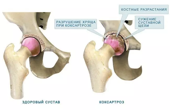
- dysplastic coxarthrosis;
- the formation of neoarthrosis;
- pathological dislocation of the hip;
- violation of posture.
Forecast
With timely diagnosis and complex treatment, the prognosis is favorable in 100% of cases. Early initiation of physiotherapy in the first weeks of life will usually ensure a complete recovery for the child.
After the completion of the correctional course, observation of an orthopedist is necessary until reaching 15-17 years
YouTube video related to the article:

Olesya Smolnyakova Therapy, clinical pharmacology and pharmacotherapy About the author
Education: higher, 2004 (GOU VPO "Kursk State Medical University"), specialty "General Medicine", qualification "Doctor". 2008-2012 - Postgraduate student of the Department of Clinical Pharmacology, KSMU, Candidate of Medical Sciences (2013, specialty "Pharmacology, Clinical Pharmacology"). 2014-2015 - professional retraining, specialty "Management in education", FSBEI HPE "KSU".
The information is generalized and provided for informational purposes only. At the first sign of illness, see your doctor. Self-medication is hazardous to health!


