- Author Rachel Wainwright wainwright@abchealthonline.com.
- Public 2023-12-15 07:39.
- Last modified 2025-11-02 20:14.
Sebaceous cyst
The content of the article:
- Kinds
- Causes and risk factors for atheroma
- Symptoms
- Complications
- Diagnostics
-
Treatment
- Conservative therapy
- Removal by minimally invasive methods
- Surgical intervention
- Prevention
- Video
A sebaceous cyst (atheroma) is a tumor-like neoplasm that appears when the sebaceous gland duct is blocked. Most people develop at least one atheroma throughout their lives.

Atheromas are usually not a cause for concern, except for a cosmetic defect.
Kinds
Cystic formations of the sebaceous glands are divided into two groups:
- True (primary atheromas) are nonvoid tumors that arise from the appendages of the epidermis.
- False cysts (retention or secondary atheromas) - appear due to thickening of sebum and / or blockage of the excretory duct of the sebaceous gland.
| View | Specifications |
| True | Slower growth is characteristic, can reach very large sizes. Such tumors are more common in females on the scalp. |
| Retention |
Faster growth is characteristic. They can be multiple and tend to merge to form lumpy conglomerates. Usually painless, may have a cyanotic hue, localized mainly in the fold behind the ear, on the cheeks, neck, on the wings of the nose, on the chest, back. On the surface of the retention cystic formation, there may be a noticeably widened opening, from which the contents of the tumor can protrude from time to time (with pressure or spontaneously). |
Causes and risk factors for atheroma
The onset of atheroma is possible in any part of the human body that has hair. However, most often this formation appears on the skin of the head, face (often localized below the mouth), neck, back, genital area.
A frequent cause of the development of a pathological process is a blockage of the sebaceous gland duct. Also, atheromas can occur with swelling of the hair follicle (for example, if it is damaged), rupture of the sebaceous gland.
Secondary atheromas often occur when the outflow of gland secretions is disturbed in patients with phlegmonous acne, oily seborrhea, and hyperhidrosis.
Risk factors include:
- hormonal disorders (for example, increased testosterone concentration);
- prolonged exposure to direct sunlight;
- frequent trauma to the skin (for example, when shaving);
- metabolic disease;
- improper use of cosmetics.
Symptoms
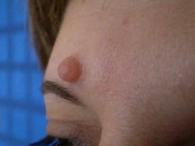
As you can see in the photo, atheroma is a densely elastic mobile formation located superficially, which has clear contours. The skin above it is not changed.
With the development of an infectious and inflammatory process, the cystic cavity can be filled with purulent contents, sometimes with an admixture of blood. The skin over the cystic formation is covered with a network of dilated capillaries; enlarged pores can be visible on the surface of the atheroma. Usually a cystic formation is 0.5-5.0 cm in diameter, but with inflammation it can increase to a large size.
With inflammation, neoplasms can be noted:
- redness and swelling of the skin over the atheroma;
- an increase in education in size;
- pain, aggravated by touching the site of the lesion;
- the release of a substance with an unpleasant odor, which may have a whitish-gray color;
- local temperature rise.
In case of inflammation and suppuration, atheroma can break out on its own. The breakthrough of an inflamed neoplasm into the subcutaneous tissue and its infection can lead to the development of an abscess and phlegmon.
Complications
If atheroma is on the scalp for a long time, it can lead to hair loss over it.
In rare cases, atheroma can become malignant, leading to the development of squamous cell skin cancer in the patient.
Diagnostics
A dermatologist's examination is usually sufficient to make a diagnosis. In doubtful cases, you may need to consult an oncologist.
When collecting complaints and anamnesis, special attention is paid to the presence of allergies in the patient, concomitant diseases (diabetes mellitus, etc.), the use of drugs that can affect the blood coagulation system.
For differential diagnosis, it may be required:
- Ultrasound - allows you to differentiate atheroma from fibroma (benign tumor from connective tissue), lipoma (neoplasm from adipose tissue), benign tumors of sweat glands;
- histological laboratory research - allows you to exclude a malignant neoplasm, Malerba epithelioma;
- X-ray examination of the bones of the skull - is carried out in the presence of large cystic formations of the sebaceous glands on the head in order to exclude a cranial hernia.

Atheromas can be large
Treatment
If the atheroma is small and does not bother the person, treatment may not be carried out. In such cases, expectant tactics are usually chosen.
Conservative therapy
In the presence of an inflammatory process, the patient can be prescribed anti-inflammatory drugs in the form of an ointment, ultra-high-frequency therapy. After the inflammation disappears, the formation is removed. In the presence of an infectious process, the patient is shown taking antibacterial drugs.
Removal by minimally invasive methods
Commonly used treatments for early atheroma include:
- Laser removal of education. It is performed under local anesthesia. When removing a growth on the head, shaving the hair is usually not required.
- Electrocoagulation. It is used in the absence of inflammation in relation to small atheromas.
- Cryodestruction. It is used in the presence of small inflamed cystic formations that are located close to the skin surface.
Surgical intervention
Surgery may be required to remove atheroma. The operation to remove the cystic formation of the sebaceous gland does not require hospitalization, it is carried out on an outpatient basis. In very rare cases, general anesthesia may be needed (for example, with a giant cystic formation), mainly the intervention is carried out under local anesthesia.
Before the operation is required:
- pass all the necessary tests;
- stop smoking and drinking alcohol per day;
- do not eat 4 hours before surgery.
In the course of surgery, according to one of the methods, the skin is cut with a scalpel (while avoiding damage to the tumor capsule), then gently press on the edges of the wound, exfoliating the atheroma, then apply cosmetic stitches and a bandage.
On the first day after surgical removal of the neoplasm, the patient may experience an increase in body temperature to subfebrile values. With a significant increase in the level of this indicator (more than 38 ° C), the development of edema and pain at the site of the operation, you should seek medical help.
Prevention
To prevent the development of cystic formations in the presence of oily skin and / or acne, it is recommended:
- Conduct professional facial cleansing with a beautician, as well as thoroughly cleanse the skin at home, following the recommendations of a specialist.
- Limit the intake of fatty foods, as well as foods high in simple carbohydrates.
- Strictly adhere to the rules of personal hygiene.
- Limit the use of decorative cosmetics, use high-quality skin care products.
Video
We offer for viewing a video on the topic of the article.

Anna Aksenova Medical journalist About the author
Education: 2004-2007 "First Kiev Medical College" specialty "Laboratory Diagnostics".
Found a mistake in the text? Select it and press Ctrl + Enter.


