- Author Rachel Wainwright wainwright@abchealthonline.com.
- Public 2024-01-15 19:51.
- Last modified 2025-11-02 20:14.
Moles in a child
The content of the article:
- Why do birthmarks appear in a child
-
Different types of birthmarks in a child
- Melanoma
- Melanomone-dangerous
- Therapeutic tactics
- Video
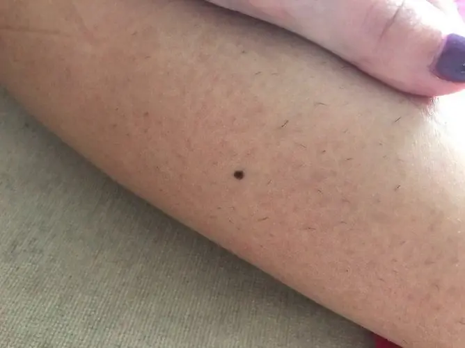
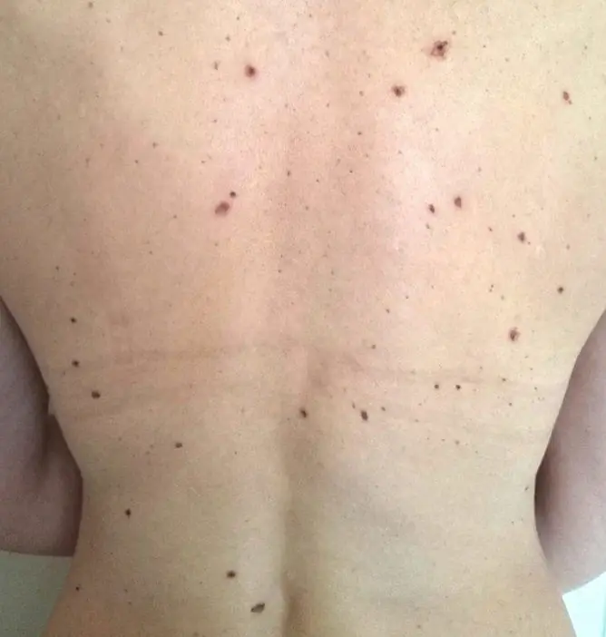
Almost everyone has one or more pigmented lesions on their skin called birthmarks, nevi, or moles. A birthmark in a child can exist from birth or appear at any age. In the first case, it is considered congenital, in the second - acquired. Most cutaneous pigmented lesions are benign and do not require special treatment, but only dynamic observation.

Moles in babies can occur for various reasons, and most of them are not dangerous
Why do birthmarks appear in a child
Nevi are formed on the skin from special pigment cells - melanocytes, which are located between the inner layer of the skin, called the dermis, and the superficial layer - the epidermis. There are several main reasons for their occurrence.
| Etiological factor | Characteristic |
| Heredity | The appearance of nevi in a specific place on the body can be programmed into a person's DNA. This is a certain defect that formed at the stage of embryonic development and is caused by a violation of the process of movement of melanoblasts - the precursors of melanocytes, from the neuroectodermal tube into the skin. The accumulation of pigment cells in the skin is the basis for the appearance of nevi. |
| Hormonal disorders | Changes in hormonal levels (puberty) and imbalance in the work of endocrine organs (stress loads, endocrine pathologies) are often factors that provoke the formation of nevi. |
| Ultraviolet spectrum radiation | One of the important functions of the melanin pigment produced by melanocytes and accumulating in them is the absorption of ultraviolet (UV) rays. Thanks to this, the deep layers of the skin are protected from radiation damage. With excessive exposure to UV rays, melanin is produced more intensively, which, in turn, provokes the formation of birthmarks. Prolonged sunbathing and frequent visits to the solarium can result in existing birthmarks growing and new ones appearing. |
| Decreased body resistance | Immunodeficiency states provoke the appearance of moles. Many formations containing pigment can appear on the skin after acute inflammatory bullous dermatoses, exposure to radiation, X-rays. Injuries, viral infections that reduce the body's defenses, contribute to the fact that new and existing nevi appear and grow. |
Different types of birthmarks in a child
All nevi in children can be divided into two large groups: dangerous in relation to the development of melanoma - a malignant tumor that develops from melanocytes, and melanomone-dangerous. The ratio of the former to the latter is 1:10.
Melanoma
| Nevus type | Description |
| Borderline pigmented (transitional, junctional) | It occurs in early childhood, usually found in the period from 6 to 12 months after the birth of the child. A distinctive feature is the location of pigment cells at the border of the dermis and epidermis. Usually it is a flat or slightly raised spot of a round or oval shape above the skin surface. Has a uniform color of various shades of brown, pink, clear and even borders, size less than 10 mm. It is localized throughout the body, including in the area of the palms, soles, and genitals. |
| Dysplastic (atypical) |
It is characterized by an increased risk of malignant degeneration due to the retention of the activity of immature melanocytes and atypia of cells of varying severity. In newborns, it is absent, it can appear in early childhood, starting from 2 years, but much more often during puberty. It looks like a spot with a separate area slightly elevated above the skin level, located in the center of the formation. The form is varied, irregular; blurred borders. The color can be black or variegated (shades of brown, black, red). Localization: frequent - on the trunk, arms, legs, dorsum of the feet, more rare - on the face. The size is usually more than 6 mm. |
| Blue (blue, Jadassona - Tiche) | Most often occurs in the period from 13 to 16 years old, girls have a 2 times more chance of forming than boys. A single formation of blue, blue or gray color less than 10 mm in diameter, clearly delimited from the surrounding skin, dense, rounded, with a smooth surface, rising in the form of a hemisphere. It can be located on any part of the body, in half of the cases it occurs on the dorsum of the hands and feet. It grows slowly, can go unnoticed for a long time. |
| Ota (Ita, dark blue orbital-maxillary) |
It belongs to congenital formations, in girls it is diagnosed 5 times more often. Usually occurs in representatives of the Mongoloid race. It is localized on the face in the area of the eye, upper jaw. Looks like a dark brown, gray-blue, or black-blue spot. May be subtle or intensely colored and disfiguring. At the birth of a child, it can be limited to the structures of the eye, then in early childhood it can appear on the skin of the face. |
| Giant pigment | Congenital melanocytic pigment formation of large sizes: in newborns and young children it occupies from 5% or more of the body surface, as the child grows, it increases and can occupy an entire anatomical region. It is a plaque of soft consistency and dark color, raised above the level of the skin, often surrounded by smaller pigmented lesions. The shape can be round or fancy, any localization. Rough hair often grows on the surface of the stain. |
Melanomone-dangerous
| Nevus type | Description |
| Intradermal pigmented | Formed in the deep layers of the skin, it is practically not found in newborns. Appears as the child grows, the amount peaks during puberty. Typical appearance: dome-shaped or warty formation, often on a stalk, 1-2 mm to several centimeters in diameter, from light shades of pink and brown to black. In girls, moles are more often found on the limbs, in boys - on the head, neck and torso. |
| Fibroepithelial | May exist from birth or appear later. It is located most often on the face or trunk, is characterized by slow growth and does not tend to change its appearance: the shape of a hemisphere, a wide base or leg, rises above the level of the skin; sizes from 2-3 mm to 1.5 cm; the consistency is soft; color - from surrounding skin to dark brown. May be injured and inflamed. |
| Papillomatous | It exists from birth or early childhood and grows slowly. It occurs anywhere on the body, often on the scalp. Has a bumpy surface, significantly protrudes above the skin surface, color - from light brown to black, sometimes covered with hair; diameter up to 6-7 cm. |
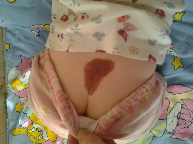
| Mongolian spot | A birthmark of a gray-bluish color, clearly defined in the region of the sacrum, buttocks, thighs, back immediately after birth. It is more common in newborns of the Mongoloid race; the appearance is associated with the accumulation of melanin in the connective tissue layer of the skin. The shape is usually irregular, sizes from 1-2 cm to 10 cm or more. As the child grows, the stain diminishes and fades, and usually disappears before puberty. |
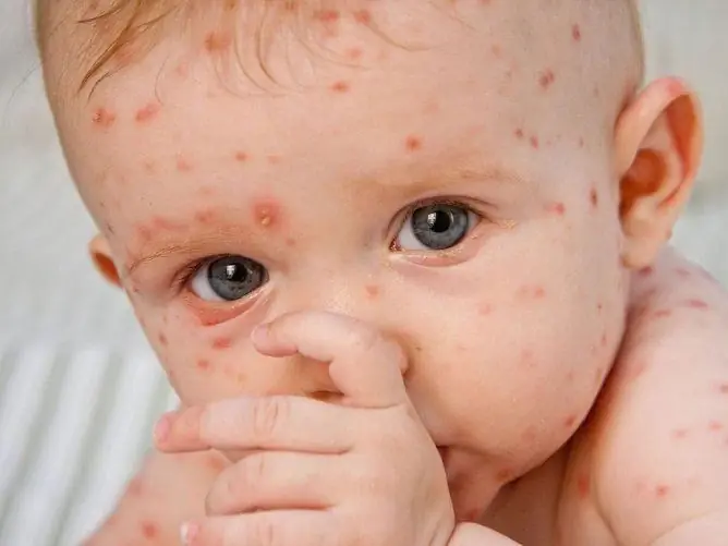
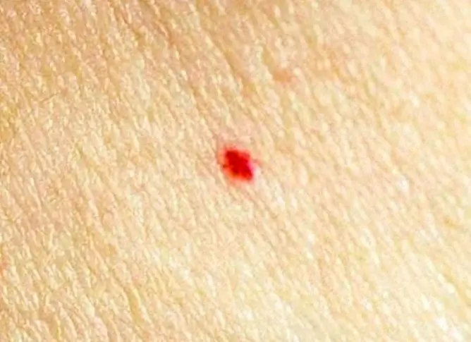
A large group of skin lesions in newborns and children are vascular moles, or hemangiomas. They arise due to the excessive growth of small vessels - capillaries. They are found immediately after birth or during the first months of life. They have clear boundaries and typical coloration: various shades of fiery red and blue-violet.
Therapeutic tactics
Most moles in a baby are safe and do not require treatment. Does this mean that parents should not be worried? It is normal for a child to have nevi, but it is necessary to know their exact origin. Treatment tactics depend on what type of nevus is diagnosed.
You also need to consult a dermatologist in the following situations:
- change in the color of education;
- rapid increase in size;
- ulceration, inflammation, bleeding;
- the appearance of itching.

If a mole is suspicious, the child should be consulted with a dermatologist.
Only a doctor, after examination and dermatoscopy, can determine what to do with the mole: observe it in dynamics or remove it. Methods for removing a nevus are varied:
- surgical excision;
- radio wave removal;
- electrocoagulation;
- cryodestruction;
- laser vaporization.
The choice of the method depends on the type of mole, its size, age of the child, localization and is determined only by a specialist after a thorough examination.
Video
We offer for viewing a video on the topic of the article.

Anna Kozlova Medical journalist About the author
Education: Rostov State Medical University, specialty "General Medicine".
Found a mistake in the text? Select it and press Ctrl + Enter.

