- Author Rachel Wainwright [email protected].
- Public 2024-01-15 19:51.
- Last modified 2025-11-02 20:14.
Osteoarthritis of the hands: causes, symptoms, treatment
The content of the article:
- Reasons for the development of DOA
-
Symptoms
- Heberden's nodules
- Bouchard's knots
- Rhizarthrosis
- Diagnostics
- Treatment of osteoarthritis of the hands
- Prevention
- Video
Osteoarthritis of the hands is one of the most common types of joint lesions. Deforming osteoarthritis (DOA) is the most common disease of articular cartilage, occurring mainly in elderly and senile people. The disease is observed in 10-15% of the total population, it is diagnosed in half of cases in people over 60 years old.

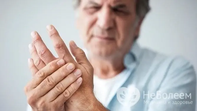
Osteoarthritis of the hands is more common in older people
Joint damage is characterized by dystrophic and degenerative changes that cause dysfunction of the cartilage, its structure and functioning. The disease ranks first among rheumatological pathologies, accounting for 60-70%. Most often, osteoarthritis affects the interphalangeal joints of the hands, hip, knee, ankle joints, etc.
Osteoarthritis of the hands is a progressive disease that inevitably leads to damage to all components of the joint: synovial membranes, synovial fluid, cartilage, subchondral areas of bones, ligaments, capsules, periarticular muscles.
One of the first symptoms is pain, which increases with the progression of the pathology, at the final stages, continuously disturbing the patient even at night. Then the deformity of the limb in the area of the joints joins, specific nodules of Heberden and Bouchard appear, morning stiffness, and motor activity is impaired.
Reasons for the development of DOA
Depending on the etiological factor, DOA is divided into primary and secondary.

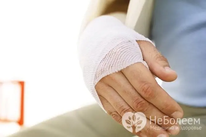
Secondary osteoarthritis can occur due to prior injuries to the wrist
In primary (cryptogenic) DOA, the causes of the onset of the disease are not fully understood, however, it is known that this form is characterized by the most frequent damage to the distal and proximal interphalangeal joints of the hands with the appearance of specific nodules of Heberden and Bouchard.
Secondary DOA develops for a number of reasons, including:
- injury, such as a dislocated or fractured wrist;
- previous surgical interventions;
- dysplasia of the joints;
- inflammatory processes in the joint (autoimmune, bacterial, parasitic or viral etiology).
These include hereditary collagen type 2 mutation, joint dysplasia, hereditary metabolic pathology or the structure of cartilage tissue, etc.
The following conditions may increase the risk of developing DOA:
- dysmetabolic and dyshormonal states that disrupt trophism, blood supply and innervation of tissues (for example, diabetes mellitus, pathologies of the thyroid and parathyroid glands, postmenopausal period, gout, hemochromatosis, etc.);
- hypothermia;
- increased load on the hands, etc.
Symptoms
Pain syndrome, signaling pathology in the joint, appears as one of the first symptoms, characterized by varying intensity and mechanism depending on the severity.
There are several types of pain in the DOA of the hands:
| Type of pain syndrome | Description |
| Mechanical pain | Most often, it occurs during exercise and subsides at rest, usually during a night's sleep. The mechanical type of pain appears one of the first and indicates a decrease in the amortization capacity of the cartilage and bone subchondral structures |
| Continuous dull night pain | It occurs due to venous stasis and an increase in intraosseous pressure. This view usually appears in the first half of the night. |
| Starting pains | Short-term, lasting no more than 15-20 minutes, occur after a period of rest and disappear during physical activity. The mechanism of this type of pain is due to the presence of an articular mouse (detritus - fragments of cartilaginous and bone destruction, deposited on the articular surface). At the first movements, detritus is pushed into the turns of the joint capsule, which is associated with the cessation of pain |
| Constant pain | It is caused by the development of reactive synovitis - aseptic inflammation of the synovium and spasm of nearby muscles |
As the disease progresses, morning stiffness, deformation of small joints, and limitation of their mobility join.
Morning stiffness is defined as the impossibility of active and passive movements in the joint, caused by inflammation of the synovial membrane, a decrease in the elasticity of the cartilage, which leads to the need for starting movements to restore motor activity. The duration of morning stiffness determines the severity of the process.
Heberden's nodules
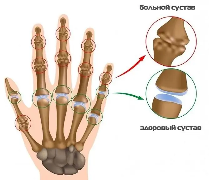
Heberden's nodules are a pathognomonic symptom for DOA of the interphalangeal joints of the hands. They are represented by marginal osteophytes, deforming the articulation, ranging in size from a grain of rice to a small pea. Nodules form on the dorsal and lateral surfaces of the distal interphalangeal joints - those that are localized closest to the nail plate.
The formation of Heberden's nodules is accompanied by a vivid clinical picture:
- characteristic pulsating pain syndrome;
- swelling and redness in the area of the affected joint.
However, one third of patients experience asymptomatic progression. In half of the cases, the onset of the disease is accompanied by a period of exacerbation, in which intense pulsating pain appears in the area of the nodules.
The skin over the nodules becomes thinner and bursts with clear fluid draining, resulting in less pain. In some cases, a breakthrough does not occur, as a result of which the pain lasts for several weeks or months, after which the symptoms subside or completely disappear, the nodules become denser and painless. Heberden's nodules inevitably lead to joint deformation and stiffness.
Bouchard's knots
Bouchard's nodules are another pathognomonic symptom for arthrosis of the fingers. They differ from Heberden's nodules in their localization and course of the process. Bouchard's nodules are formed in the region of the median interphalangeal joints (the second from the nail plate) and affect their lateral surfaces. This leads to the formation of a specific fusiform shape of the fingers involved in the pathological process.

Gradual deformation of the joints leads to severe symptoms
According to the clinical course, Bouchard's nodules differ from Heberden's nodules in less pronounced symptoms. Progression occurs gradually with mild pain, but the process also leads to deformation and stiffness of the joint.
Rhizarthrosis
With polyosteoarthritis, the joints of the thumb may be involved. Osteoarthritis of the thumb is called rhizarthrosis.
Diagnostics
Diagnostics is based on the clinical picture, patient complaints, and the results of laboratory and instrumental studies.

Instrumental studies are carried out to confirm the diagnosis.
There are three diagnostic criteria for the diagnosis of hand osteoarthritis:
- pain, stiffness, or stiffness in your hands for the past month;
- dense thickening of two or more joints (II and III distal interphalangeal, II and III proximal interphalangeal, carpometacarpal joints of both hands);
- the number of edematous metacarpophalangeal joints is less than three.
Perhaps the appointment of an X-ray examination, computed and magnetic resonance imaging (the last two diagnostic methods are used extremely rarely).
X-ray of the hand visualizes the state of bone structures, joint space, and the presence of osteophytes. An X-ray image allows you to indirectly assess the condition of the cartilage.
Treatment of osteoarthritis of the hands
Treatment is carried out on an outpatient basis, comprehensively, conservatively.
Physiotherapeutic methods, physiotherapy exercises, pharmacological agents, diet therapy are used. Traditional methods of treatment are rarely used due to their ineffectiveness.
| Treatment type | Methods |
| Physiotherapy | Phonophoresis, iontophoresis, balneotherapy, sulfide, radon baths, light therapy, electromyostimulation, ultrasound therapy, diathermy, cryotherapy, exercise therapy (therapeutic exercises), massage |
| Diet therapy | Diet aimed at weight loss, normalization of endocrine and metabolic disorders |
| Pharmacotherapy |
NSAIDs - non-steroidal anti-inflammatory drugs - are used to reduce pain, swelling. Paracetamol, diclofenac, ibuprofen, nimesulide, ketorolac, meloxicam, celecoxib, etc. are prescribed. Chondroprotectors - used in the early stages (chondroitin sulfate, glucosamine). The action of the drugs is aimed at restoring damaged cartilage |
| Intra-articular injections |
The introduction of glucocorticoids (hormonal drugs): reduces the severity of intra-articular inflammation and reduces pain. It is known that glucocorticoids negatively affect cartilage, therefore, the method is used in extreme cases when NSAIDs do not show their effect sufficiently Hyaluronic acid injection: facilitates the process |
Prevention
There is no specific prevention of osteoarthritis of the hands.
Recommended:
- dose physical activity, avoiding prolonged static and mechanical overload of the joints;
- try to avoid injury;
- timely diagnose and correct congenital anomalies of the musculoskeletal system;
- normalize excess body weight.
Video
We offer for viewing a video on the topic of the article.

Anna Kozlova Medical journalist About the author
Education: Rostov State Medical University, specialty "General Medicine".
Found a mistake in the text? Select it and press Ctrl + Enter.


