- Author Rachel Wainwright wainwright@abchealthonline.com.
- Public 2024-01-15 19:51.
- Last modified 2025-11-02 20:14.
Breast cyst
The content of the article:
- Classification
- Reasons for the appearance
-
Disease symptoms
- Solitary cyst
- Ductal cyst (intraductal papilloma)
- Diffuse forms with a predominance of the cystic component (Reclus disease, adenomatosis)
- Diffuse forms with a predominance of the fibrous component
- Cystic fibrous form
- Other forms
-
Treatment
- Drug therapy
- Surgery
- Folk remedies
- Video
What is a breast cyst? This type of neoplasm, according to the histological classification of benign tumors by WHO (2012), belongs to the group of fibrocystic diseases of the mammary glands (mastopathy ICD 10 code N60.0). There is no separate category of breast cyst, it is considered as a manifestation of cystic fibrous disease.
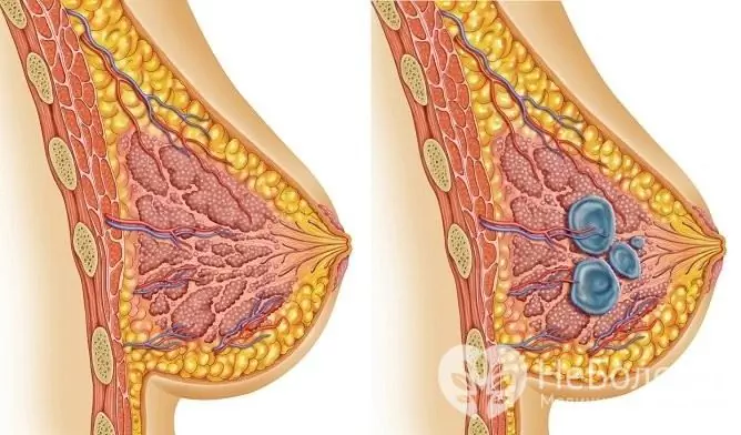
A breast cyst is one of the manifestations of fibrocystic breast disease.
Classification
There is the following classification of mastopathy (N. I. Rozhkova, 1983):
| View | Characteristic | Options |
| Diffuse form of fibrocystic mastopathy |
Multiple small cysts or overgrowth of connective tissue. 70% of women have a mixed form. |
1. With a predominance of the cystic component (Reclus disease, adenomatosis). 2. With a predominance of the fibrous component. 3. With a predominance of the glandular component (there is no cystic cavity, and is not considered in this section). 4. Fibrocystic form. 5. Sclerosing adenosis (there is no cystic cavity, and is not considered in this section). |
| Nodular form of fibrocystic mastopathy |
Depending on the options for the course, the pathology is a single cyst or an overgrowth of connective tissue, which leads to the appearance of cysts. |
1. Solitary cyst. 2. Ductal cyst of the mammary gland (intraductal papilloma). 3. Fibroadenoma (there is no cystic cavity, and is not considered in this section). |
Depending on the location and structure, the following forms are distinguished:
- lobular;
- ductal;
- fibrous;
- cystic.
Depending on the degree of proliferation:
- non-proliferative form - no signs of malignancy;
- with moderately pronounced intraductal proliferation - precancer;
- with atypical intraductal proliferation - cancer.
Appears more often in women during menopause. After 60 years, more often the risk of nodular forms of mastopathy doubles.
Reasons for the appearance
The reasons for the development of benign neoplasms are in many ways similar to the causes of malignant forms. Formations are related to polyetiological diseases.
Risk factors for cysts:
- Late term of the first birth (over 35 years old) and a large number of abortions in history.
- Low birth rate. The break between childbirth is more than 5-7 years.
- The birth of a large fetus (weight over 5 kg).
- No or short period of breastfeeding. According to WHO, breastfeeding should be carried out up to 3 years.
- Disruption of the ovaries and, as a result, a violation of the menstrual cycle (dysmenorrhea, amenorrhea).
- Inflammatory diseases of the pelvic organs. Of particular importance are diseases of the uterine appendages (ovaries and fallopian tubes).
- Disorders in the hormonal system. Of particular importance are the concomitant hormonal diseases of the reproductive system (endometriosis, fibroids). Systemic disorders of the endocrine system (diabetes mellitus, thyroid pathology, dysfunction of the adrenal cortex) are also important.
- Benign and malignant ovarian tumors.
- Metabolic disorders (in particular, liver disease).
- Genetic risk factors. Mutation of some genes (BRCA 1,2) does not cause tumor development, but leads to incorrect functioning of the cell genome, being a predisposing component.
- External factors (stress, unhealthy diet).
The pathogenesis is based on a violation of the hypothalamic-pituitary-ovarian system (hormone-conditioned pathology):
- insufficient effect of progesterone - reduces the level of estrogens and their effect on breast tissue, ensures cell differentiation, prevents cells from dividing uncontrollably;
- excess estradiol - enhances the division of breast cells, lead to obstruction of the ducts and the development of cysts;
- violation of the mechanism of apoptosis (natural cell death).
Spontaneously, cystic cavities rarely resolve.
Disease symptoms
In each case, the symptoms will manifest themselves differently depending on the size, location, time of onset, and the involvement of surrounding tissues.
The main symptom complexes:
- pain syndrome;
- intoxication syndrome;
- local changes.
Solitary cyst
It is a cavity with liquid contents, which is located in a capsule, the breast tissue is not changed. The fluid is serous in nature, less often hemorrhagic or purulent. The cavity is filled and formed gradually, so the clinical picture does not develop immediately. The size varies:
- microcyst - up to 10 mm;
- small - up to 3 cm;
- medium - 3-5 cm;
- large - more than 5 cm.
Features of education:
- mobile, not soldered to the surrounding tissues;
- rounded;
- single;
- not related to the skin;
- single-chamber (less often it is two-chamber).
The main symptoms are:
- There are often no symptoms. Accidental detection during routine examinations (palpation or ultrasound).
- Pain (mastalgia) of varying severity. On palpation, the doctor may detect painful tension in the breast (mastodynia). It has a clear relationship with the menstrual cycle (the gland begins to hurt a lot 10-14 days before menstruation, after which the pain almost completely disappears). More often the pain syndrome is cyclical, but with concomitant ovarian dysfunction it is permanent.
- Discharge from the nipples is extremely rare. By nature, transparent, without signs of suppuration.
- The appearance of the mammary gland is not changed. As the cyst grows, slight asymmetry may occur.
Treatment tactics: expectant nature (in the absence of growth, an ultrasound scan and mammography are required every six months). The forecast is favorable.
Ductal cyst (intraductal papilloma)
It is formed as a result of the proliferation of the epithelium inside the dilated excretory duct of the gland (the cyst is not represented by a typical formation with a capsule, but by the expanded walls of the duct). Due to the increase in the number of collagen fibers, the normal drainage of the lobules of the mammary gland is disturbed, stagnation and a gradual increase in their volume occurs (a pseocyst is formed).
Features:
- location more often in only one duct, less often in several;
- localization in the subareolar zone or in the nipple.
It is impossible to palpate the tumor itself (treelike growth in the duct of the gland). On examination, only dilated lobules can be found, which are defined as slightly painful formations and can cause difficulties in making a diagnosis (differential diagnosis with other forms of tumor-like formations is required).
Clinical picture:
- Profuse serous, bloody, or hemorrhagic nipple discharge. The appearance of discharge on linen after sleep, after taking a bath, on clothes is typical.
- This type of formation does not cause pain, it can only cause discomfort in the breast area.
- Locally, slight redness in the areola area can be determined (there is no asymmetry of the breast on examination or in the photo).
- The skin is not involved, so there is no retraction or protrusion (the lemon peel symptom is negative).
Patients with suspected ductal cysts should be evaluated with MRI / CT. Why is this necessary? The fact is that this form refers to precancerous conditions.
Diffuse forms with a predominance of the cystic component (Reclus disease, adenomatosis)
Since it belongs to diffuse mastopathy, it is characterized by multiple cysts of different diameters and localization (often multiple small cystic cavities). The cystic (fluid) component predominates, and the connective tissue cords in the interlobular space are weakly expressed.
Features of cystic formations:
- multiple, elastic;
- insignificant mobility;
- the size varies widely.
Clinical picture:
- Severe pain syndrome. Has an addiction to the menstrual cycle.
- Discharge from the nipple of a different nature (serous, purulent), which occurs when pressing on the nipple. The amount is variable.
- Inflammation of regional lymph nodes (in particular, in the armpit).
- Densely elastic round formations are determined by palpation. There are no external signs in 70% of cases. With the growth of foci, skin hyperemia or lemon peel syndrome can be observed.
- Often there is a kind of paraneoplastic syndrome (headaches, edema, dyspeptic symptoms).
Diffuse forms with a predominance of the fibrous component
The form is similar to the previous one, also represented by multiple cysts. The difference lies in the ratio of liquid and tissue components. Features:
- dense on palpation;
- soldered to surrounding tissues;
- motionless or poorly mobile;
- sometimes the skin is affected (pathological retraction or protrusion).
The clinical manifestations are the same for the two forms, but in the case of the fibrous one they are somewhat stronger. The prognosis is relatively favorable (the form is a variant of a precancerous condition).

In some cases, surgical removal of the breast cyst is required
Cystic fibrous form
Combined type, represented by multiple cysts surrounded by dense connective tissue structures (nodes, plexuses). Normal breast tissue is almost completely reborn.
Features:
- multiple;
- elongated flat cake shape;
- lobular structure (connective tissue squeezes cysts, separating them);
- poorly mobile (not associated with the chest wall and skin).
Clinical picture:
- Pain syndrome associated with menstruation. The feeling of fullness is replaced by a feeling of pain as menstruation approaches, then it disappears for a short period of time. Has a diffuse nature (the predominance of pain in the upper-outer quadrants). It can radiate to the arm, axillary zone, nipple (if localized next to one of the ducts).
- On palpation, the mammary gland resembles a cobblestone pavement (multiple painful lobules). Pronounced asymmetry in relation to a healthy gland, lemon peel over protrusion sites. Visual heterogeneity of the gland (tuberosity).
- There is no spontaneous nipple discharge. When pressed, they can have a different character and color (transparent, serous, turbid-serous, greenish). Rarely, purulent discharge (thick, yellowish-greenish) may occur, but cytological examination of signs of inflammation does not reveal. It is dangerous to have a discharge with an admixture of blood, since the question arises about a malignant process.
- A pronounced paraneoplastic syndrome is characteristic.
The course is complicated by the psychoemotional state (depression, the stage of denial). Consultation of a psychotherapist is shown.
Other forms
There are several forms of cysts that do not belong to mastopathy and act as complications of the underlying disease:
- Post-traumatic injury. Cysts occur as a result of a blow and often contain hemorrhagic contents. Under normal conditions, they pass on their own. In case of infection, suppuration is possible. There are no long term consequences.
- Milk cyst (galactocele). It occurs due to a violation of the outflow of milk (incorrect use of the breast pump, incorrect feeding technique). The difference between this type of cyst and ductal cysts is that there is no mechanical barrier in the form of papilloma inside the duct. No treatment is required, since 80% of it goes away on its own. In case of infection, suppuration is possible.
- Polycystic. In this case, normal breast tissue is absent, it is completely replaced by cysts of different sizes. The disease is congenital and does not belong to tumor-like processes.
The prognosis for all these types is favorable. Patients usually recover within a month; repeated courses of therapy are rarely needed.
Treatment
What to do if mastopathy is found? Treatment of a cyst in the chest depends on the characteristics of the process, and is carried out in two main ways:
- Conservative therapy with drugs and constant monitoring of growth (ultrasound, mammography).
- Surgical treatment is an operation in a planned manner.
Drug therapy
| Type of therapy | Group of drugs | Example |
| Non-hormonal drugs | Phytotherapy | Mastodinon, Indinol |
| Vitamin therapy | Vitamin E, Ascorutin | |
| Hormonal drugs | Estrogens (replacement therapy). | Femoston |
| Progesterone drugs (replacement therapy) | Utrozhestan, Duphaston | |
| Selective estrogen receptor modulator | Tamoxifen, Fareston | |
| Antiprolactins | Bromocriptine, Parlodel, Dostinex | |
| Inhibitors of the pituitary gonadotropic function. | Goserelin, Buserelin | |
| Symptomatic drugs | Hepatoprotectors, choleretic drugs | Essentiale, Karsil |
| Sedatives | Gelarium | |
| Diuretic drugs | Veroshpiron, Furosemide | |
| Immunomodulators (anti-inflammatory, anti-edema, anti-proliferative action) | Cycloferon, Amiksin. | |
| Antiprostaglandins (relieve premenstrual syndrome and breast edema) | Naproxen, Nimesulide. |
Each form of cystic formations requires a strictly individual selection of combinations of drugs and dosages, therefore, only the attending physician should prescribe them.
Surgery
Surgical treatment methods:
- puncture;
- sclerotherapy (rarely and in the elderly);
- enucleation (exfoliation of the cyst);
- sectoral resection of the gland.
Surgical removal of a neoplasm is indicated after the lack of effect from conservative therapy, with large neoplasms or when they grow into adjacent tissues.
Folk remedies
Treatment of breast cysts with traditional medicine is ineffective.
Video
We offer for viewing a video on the topic of the article.

Anna Kozlova Medical journalist About the author
Education: Rostov State Medical University, specialty "General Medicine".
The information is generalized and provided for informational purposes only. At the first sign of illness, see your doctor. Self-medication is hazardous to health!


