- Author Rachel Wainwright wainwright@abchealthonline.com.
- Public 2023-12-15 07:39.
- Last modified 2025-11-02 20:14.
Radius
The radius is a paired bone that is part of the forearm and is located outward and anterior to the ulna.

The structure of the radius
The radius is distinguished:
- The body is triangular in shape with three edges (anterior, interosseous and posterior) and three surfaces (anterior, posterior, and lateral).
- Upper and lower epiphysis.
The anterior surface has a somewhat concave shape and contains a feeding opening, which begins the feeding channel.
The smooth posterior surface is separated from the lateral surface by the posterior edge.
Muscle tendons lie in the grooves of the posterior surface of the lower epiphysis of the radius.
The carpal articular surface is the junction of the lower surface of the radius with the bones of the wrist.
Fracture of the radius
Fracture of the radius at the "typical site" - the most common among the total number of different fractures of the forearm bones. Falling onto an outstretched arm is usually the cause of this injury. In case of injury, in addition to a fracture, such concomitant injuries as:
- Dislocation of the lunate bone;
- Fracture of the scaphoid and styloid process;
- Ligament ruptures (wrist and radiator).
Most often, older people, especially women, are prone to fractures, which is associated with metabolic disorders and the development of osteoporosis.
Symptoms of a radius fracture are:
- Sharp pain;
- "Fork-shaped" curvature of the forearm;
- Dysfunction of the hand and fingers;
- Edema.
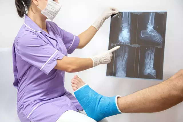
In order to establish whether the radius has been displaced during a fracture, an X-ray should be taken in two projections.
If necessary, the traumatologist sets the displaced bone fragments, after which a plaster splint is applied.
Control X-rays are usually taken 10-12 days after the injury (after the edema subsides). Sometimes, after reducing the edema, the limb is not fixed with a sufficiently high quality plaster cast, which can subsequently lead to secondary displacement of the debris.
With a fracture without displacement of the radius, the period of immobilization (rest) is four to five weeks. If the fracture was accompanied by displacement, the cast is applied for up to eight weeks.
One of the main complications of radial displacement is post-traumatic dystrophy of the hand (otherwise, Turner's trophoneurosis). It can be caused by too closely applied plaster cast, which usually occurs due to the increase in edema for several days after the injury.
In some cases, with unstable fractures of the radius, which tend to secondary displacement of fragments, an operation is required to prevent the development of arm deformities and nerve compression. In some cases, osteotomy is performed with replacement of the defect with artificial or own bone. Typically, the plate is removed seven months after the arm is fully functional and the bone is fully restored.
Rehabilitation after a fracture of the radius
It is recommended to start rehabilitation after a fracture of the radius as soon as possible (immediately after the pain subsides). It is desirable to carry out a comprehensive recovery, which would include not only therapeutic gymnastics, but also massage, the use of warming ointments and compresses, and physiotherapy. In addition, some exercises are recommended to be carried out in warm water to relieve stress.
Exercise should cover all free joints of the injured limb. You should especially pay attention to the warm-up of the fingers: they should be unclenched and squeezed, as well as collect various small objects (buttons, matches).
The entire exercise cycle should be carried out for at least half an hour, two to three times a day.
The recovery process after a fracture is facilitated by massage procedures using special ointments and gels:
- Activating metabolism at the site of application;
- Relieve inflammation;
- Accelerating healing;
- Pain relief.
Timely rehabilitation after a fracture of the radial bone contributes not only to the fastest restoration of the functional capabilities of the hand, but also prevents the development of trophoneurosis.
Found a mistake in the text? Select it and press Ctrl + Enter.


