- Author Rachel Wainwright wainwright@abchealthonline.com.
- Public 2023-12-15 07:39.
- Last modified 2025-11-02 20:14.
Endoscopy

Endoscopy - diagnostics of internal organs using special devices - endoscopes.
Endoscopy method
The method of endoscopic examination is that a soft tube is inserted through the holes into the human body, at the end of which a lighting device and a microcamera are attached. This tube is called an endoscope. Its diameter does not exceed 4 mm.
Different endoscopes are designed for different areas of medicine. For endoscopy of the stomach, upper digestive tract, duodenum, gastroduodenoscopes are used, enteroscopes are used to examine the small intestine, colonoscopes are used for intestinal endoscopy, and bronchoscopes are used for the respiratory tract.
With some manipulations, the endoscope is inserted through the mouth (gastric endoscopy), while others through the rectum (intestinal endoscopy), through the larynx, urethra and nose (nasopharyngeal endoscopy). To carry out, for example, laparoscopy, special holes have to be made in the abdominal cavity.
Kinds
There are many types of endoscopic examination. Using this procedure, you can study the state of such vital organs as the abdominal cavity, vagina, small and 12 duodenum, ureters, bile ducts, esophagus, hearing organs, bronchi, uterine cavity, as well as make gastric endoscopy, intestinal endoscopy, endoscopy nasopharynx.
The endoscope can be passed through the vessels and check their condition, as well as view the heart and cardiac chambers. In our age, an endoscope can even get into the brain and give the doctor the opportunity to view the ventricles of the brain.
All types of endoscopic studies are aimed at identifying minimal changes in the mucous membrane, which in the future can lead to oncology. Also, the procedure allows you to identify oncology at an early stage and remove the tumor, which significantly increases the chances of cancer patients for survival.
Cancer in the early stages cannot be detected at all with the help of another study, so today there is no alternative to endoscopy.
In addition to diagnostics, this procedure has found wide application in surgery, urology, gynecology and other fields. With its help, doctors stop bleeding, remove tumors in the early stages. The procedure allows not only to diagnose internal organs, but also to take a tissue sample of the neoplasm for analysis.
The technique is also widely used in plastic surgery, for example, forehead and eyebrow endoscopy. Forehead endoscopy allows you to raise the eyebrows, remove or reduce the number of expression lines on the forehead and between the eyebrows. Forehead endoscopy is very popular due to the fact that it leaves almost no scars.
How is the endoscopy procedure performed?
During gastric endoscopy, the apparatus is inserted through the mouth and the mucous membrane is examined on the monitor. In this case, air is supplied through the endoscope - this is necessary for a more detailed examination. The procedure takes about 15-20 minutes.

For the study to be more accurate, it is necessary to properly prepare for it. 8-12 hours before the procedure, it is advisable not to eat or drink anything.
Gastroscopy is a painful examination that causes the patient to gag.
Transnasal endoscopy is tolerated by patients much easier, since the gag reflex is absent.
Gastric endoscopy is done to clarify the diagnosis and identify changes.
Bowel endoscopy is more painful and time consuming. Pain can be caused by features of the intestine, adhesions. The procedure itself takes from 30 minutes to 1 hour. Anesthesia is often used during colonoscopy.
Preparation is also important when performing a colonoscopy. Here it is recommended to switch to a slag-free diet three days before the procedure.
Indications for colonoscopy are stool disorders, mucus and blood discharge, painful sensations, bleeding from the colon.
Bronchoscopy is performed by inserting a thin endoscope through the nose, larynx, and vocal cords directly into the trachea. This allows you to view the bronchial tree from the inside. The study is indicated for pneumonia, bronchitis, suspected tumor.
During endoscopy of the nasopharynx, an endoscope is inserted into the nose, which allows you to see the picture inside the nose and possible polyps. Endoscopy of the nasopharynx is indicated for shortness of breath, nosebleeds, impaired sense of smell, polyps and vague headaches.
Endoscopy of the nasopharynx allows you to identify pathological changes in the nasal mucosa without the intervention of surgical methods.
Video capsule endoscopy
This type is a new direction in medicine. The method consists in the fact that the patient swallows a plastic capsule, which is no larger than a regular medicine capsule. The capsule passes through all the digestive organs, while the entire image is recorded on a special apparatus, and he, in turn, transmits all data to the screen.
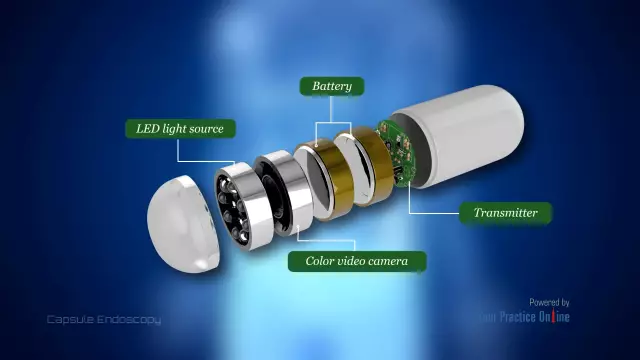
Video capsule endoscopy was patented in America at the beginning of this century and is rapidly gaining momentum. The capsule itself weighs 4 grams, and its length is 2.5 cm. One end of the capsule is transparent, behind it is a lens, a micro-camera and LEDs. The rest of the capsule contains the transmitter, battery, and antenna.
Video capsule endoscopy is very convenient, as it allows without interrupting the patient's main activities to carry out a complete endoscopy of the stomach, endoscopy of the intestines and the digestive tract. In addition, such a study allows you to see even those sections of the intestine that are not accessible during a conventional endoscopic examination.
However, video capsule endoscopy has one significant drawback. Unfortunately, this technique can only be used to study the digestive system.
Found a mistake in the text? Select it and press Ctrl + Enter.


