- Author Rachel Wainwright wainwright@abchealthonline.com.
- Public 2023-12-15 07:39.
- Last modified 2025-11-02 20:14.
Effusion
Effusion is a pathological condition characterized by the accumulation of any biological fluid in one of the body cavities. Effusion is a symptom of inflammation or excess blood in internal organs or tissues, and treatment should be directed towards the underlying disease.
Pleural effusion

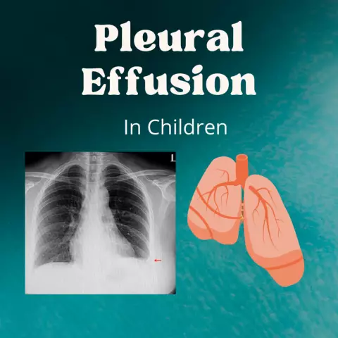
Pleural effusion is accompanied by chest pain and shortness of breath. This is the accumulation of the following fluids in the pleural cavity:
- Blood. Hemothorax can occur due to chest trauma, aortic aneurysm, or bleeding disorders;
- Pus. Empyema is a complication of pneumonia, esophageal rupture, abdominal abscess, or surgery;
- Lymphs. Chylothorax occurs with various damage to the main lymphatic duct or when it is blocked by a tumor.
Diseases that can provoke pleural effusion:
- Chest trauma;
- Heart surgery;
- Tumors;
- Heart failure;
- Pulmonary embolism;
- Pneumonia;
- Tuberculosis;
- Histoplasmosis;
- Cryptococcosis;
- Blastomycosis;
- Coccioidomycosis;
- Pancreatitis;
- Cirrhosis of the liver;
- Rheumatoid arthritis;
- Abscess under the diaphragm;
- Systemic lupus erythematosus;
- Low blood protein.
The cause of the effusion can be the incorrect introduction of intravenous catheters or plastic tubes, as well as some drugs - hydralazine, procainamide, isoniazid, phenytoin, and others.
Diagnostics of the accumulation of fluids in the pleural cavity includes:
- Chest X-ray - reveals fluid;
- Computed tomography - confirms the results of an X-ray, detects lung abscess, pneumonia or tumor;
- Ultrasound - identifies the location of the smallest amount of pleural fluid, making it easier for the doctor to drain it.
In cases where the source of pleural effusion could not be identified during these procedures, puncture, biopsy, or bronchoscopy are performed. Puncture allows you to establish the presence of fungi, bacteria and malignant cells in the chemical composition of the liquid. For this, the liquid is separated with a needle, while draining the cavity. If there is not enough sample for diagnosis, then an open pleural biopsy is done - the chest is slightly cut and tissue is taken using a thoratoscope. Sometimes they resort to bronchoscopy - a direct visual study of the airways through a bronchoscope. According to statistics, in 20% of cases, the cause of the effusion cannot be determined.
Depending on the severity of the effusion, excess fluid is removed with a needle, catheter, or plastic tube connected to the drainage system.
Knee effusion
Knee effusion is usually caused by inflammation, injury, or overuse. To determine the cause of the accumulation of fluid in the knee, you need to examine a sample of it. Factors such as age, obesity, and occupation contribute to joint effusion. The risk increases after age 55, as the elderly are more likely to develop joint diseases. Additional stress on the knees can occur in people who are overweight or in professional athletes. Over time, overloading will damage the cartilage, which is a common cause of effusion.
Fluid can accumulate after bone fractures, meniscus or ligament rupture, as well as in chronic diseases:
- Improper blood clotting;
- Tumors;
- Kist;
- Osteoarthritis;
- Bursitis;
- Rheumatoid arthritis;
- Septic arthritis;
- Gout;
- Pseudogout.
Signs of a joint effusion are:
- Puffiness. Around the kneecap, tissue swelling is observed, which is especially noticeable when comparing a sick and healthy knee;
- Stiffness. Excessive fluid prevents the joint from moving freely and does not allow the leg to be fully extended;
- Pain. The effusion can restrict movement. Sometimes the pain leads to the fact that a person cannot stand up.
Diagnostics consists in carrying out a number of procedures:
- X-ray. Shows signs of arthritis, joint destruction, bone fractures;
- Ultrasound. Reveals diseases of tendons and ligaments;
- Magnetic resonance imaging. Identifies minor damage to joints and tissues.
Invasive techniques include blood tests, joint aspiration, and arthroscopy. A blood test can diagnose infectious and inflammatory conditions that accompany an effusion, such as Lyme disease or gout. Joint aspiration, or arthrocentesis, is done to check for bacteria, blood, uric acid crystals and other impurities in the accumulated fluid. Samples can also be taken while examining the joint surface with an arthroscope inserted into the joint.
Medical treatment for knee effusion is intended to address the underlying cause, and in severe cases, surgery may involve removing fluid or replacing the joint.
Pelvic effusion
Pelvic effusion is a build-up of free fluid that can be normal. For example, in women, immediately after ovulation, the liquid content from the ruptured follicle enters the space behind the uterus, and disappears after 2-3 days. This sign is used in the treatment of infertility as a kind of ovulation marker.

The causes of effusion can be the following pathologies:
- Liver disease;
- Endometriosis;
- Ovarian cyst rupture;
- Purulent salpingitis;
- Ectopic pregnancy;
- Malignant tumor;
- Bleeding in the abdominal cavity of any origin.
Diagnostics is carried out mainly using ultrasound. Treatment can be conservative or operative, depending on the associated diseases.
The presence of effusion in the joint, pleural cavity, or pelvic area is an important diagnostic symptom of various inflammatory diseases and requires immediate medical attention.
The information is generalized and provided for informational purposes only. At the first sign of illness, see your doctor. Self-medication is hazardous to health!


