- Author Rachel Wainwright wainwright@abchealthonline.com.
- Public 2023-12-15 07:39.
- Last modified 2025-11-02 20:14.
Ventriculomegaly

Ventriculomegaly is a fetal abnormality in which there is a small or significant increase in the size of the ventricles of the brain, which leads to neurological disorders and diseases of the brain.
Causes and symptoms of ventriculomegaly
Pathological ventriculomegaly in the fetus can be either isolated or a defect associated with other developmental pathologies. With this disease, the fetus has a large size of the ventricles of the brain, reaching up to 12-20 mm.
In modern medicine, among the main causes of ventriculomegaly, chromosomal abnormalities are distinguished, which are observed in 17-20% of women with pathologies during pregnancy.
Ventriculomegaly can be caused by obstructive hydrocephalus, physical injury, infectious disease, hemorrhage, and hereditary factors. Pathology is enhanced by the presence of other developmental anomalies.
Fetal ventriculomegaly can lead to the development of Down syndrome, Turner and Edwards syndrome in a child. This disease affects the changes in the structures of the brain, heart and musculoskeletal system.
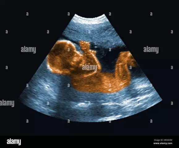
Symptoms of ventriculomegaly can be clearly identified with ultrasound, and their appearance is noticeable at 20-23 weeks of gestation. In some cases, the pathology is recorded at the beginning of the third trimester of pregnancy, the best period for diagnosing and detecting symptoms of the disease is 25-26 weeks.
If ventriculomegaly is a single fetal pathology, then the likelihood of severe chromosomal abnormalities is low. The attending physician determines the risk of developing brain diseases in a child in accordance with the increase in the width of the ventricles.
Medical studies have found that the risk of ventriculomegaly is high in pregnant women over the age of 35 (the incidence of diseases is 0.5-0.7%), and in young women in labor it is significantly reduced.
In pediatrics, there are three main types of ventriculomegaly:
- severe type with the presence of large increases in the ventricles of the fetal or newborn brain, as well as combined with other brain pathologies;
- middle type with an increase in ventricles up to 15 mm and small changes in the outflow of cerebrospinal fluid;
- mild type, which has a single character and does not require serious treatment.
Diagnosis of the disease
Ventriculomegaly is diagnosed during pregnancy (from 17 to 33 weeks) using ultrasound and spectral karyotyping of the fetus. Perinatal examinations should include examination of all anatomical structures of the fetus, especially the ventricular system of the brain.

To establish an accurate diagnosis, a transverse scan of the fetal head is performed to determine the threshold value of the lateral ventricles of the brain. Ventriculomegaly is defined when the ventricles are larger than 10 mm.
Treatment of ventriculomegaly
The main treatment of ventriculomegaly is aimed at preventing the onset of the consequences of this pathology, which can be severe diseases of the brain and central nervous system.
A pediatrician and a neurologist prescribe antihypoxants, diuretics and vitamins as drug therapy.
For the treatment of ventriculomegaly, massage and physiotherapy exercises (static exercises with loads on the pelvic muscles and pelvic floor) are prescribed.
As drugs intended to prevent the development of neurological disorders in a child, drugs are prescribed to retain potassium in the body.
The information is generalized and provided for informational purposes only. At the first sign of illness, see your doctor. Self-medication is hazardous to health!


