- Author Rachel Wainwright wainwright@abchealthonline.com.
- Public 2023-12-15 07:39.
- Last modified 2025-11-02 20:14.
Fetal ultrasound

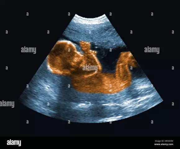
Ultrasound (ultrasound) of the fetus is the main diagnostic method during pregnancy. How many times a pregnant woman should do an ultrasound of the fetus is determined by the gynecologist who is observing her. The goals of an ultrasound scan of the fetus differ, depending on which period of pregnancy it is performed at.
Usually, the number of scheduled (mandatory) ultrasounds does not exceed 5 times:
1. To determine the pregnancy itself - approximately for a period of 5 - 7 weeks;
2. To assess the development of the fetus inside the womb, as well as the state of the mother's placenta and exclude developmental defects. An ultrasound of the fetal heart is performed - for a period of 11 - 13 weeks;
3. To exclude malformations, assess the state of the placenta and amniotic fluid in it, as well as to determine the sex of the child. The size of the fetus is necessarily determined by ultrasound and ultrasound of the fetal heart - for a period of 19 - 21 weeks;
4. To determine the approximate weight of the child and the condition of the umbilical cord, as well as the commensurability of the size of his head and the birth canal of the mother. The size of the fetus is determined by ultrasound - for a period of 32 - 34 weeks;
5. To prepare for childbirth, in order to anticipate possible complications - immediately before childbirth, with the first contractions or when the amniotic fluid flows.
The main types of fetal ultrasound and how they are performed
There are such basic methods of fetal ultrasound as:
1. Transabdominal (the sensor is located on the woman's abdomen);
2. Transvaginal (the sensor is inserted into the vagina).
Both types of procedures are completely painless for a woman, and ultrasound is not harmful to the fetus.
Transvaginal ultrasound is more accurate. Recently, three-dimensional and four-dimensional ultrasound of the fetus are considered very common ways to obtain additional information about pregnancy.

For three-dimensional or 3D ultrasound, a complex computer program is used that provides a spatial image of the fetus, which is based on two-dimensional (flat) images. Basically, a 3D ultrasound scan produces an accurate photograph of the fetus. Currently, this diagnostic method is used for the early detection of fetal malformations that could be missed during a routine ultrasound scan. For example, using an ultrasound of the fetal heart, it is possible to identify defects in the development of the organ.
Four-dimensional or 4D-ultrasound of the fetus allows you to see the volumetric image of the child in real time, while his movements and the work of all internal organs are visible.
Expectant mothers are often worried about whether ultrasound is harmful to the fetus. So, modern ultrasound machines are completely harmless for the mother and the unborn baby. In addition, they allow not only to determine the size of the fetus by ultrasound, to make an ultrasound of the fetal heart, but also to print photographs, and also record a video.
An ultrasound Doppler is used to conduct an ultrasound of the fetal heart. This device makes it possible to study the blood circulation in the blood vessels, in the heart of the child and in the umbilical cord, as well as in the vessels of the mother's placenta. Doppler ultrasound data are essential for early detection of potential child health problems:
- Heart defects;
- Blood vessel abnormalities;
- Placenta problems.
Doppler ultrasound is recommended by doctors for all pregnant women at 12 and (or) 20 weeks of gestation.
An unscheduled ultrasound scan may be prescribed by a gynecologist in the following cases:
1. Bloody discharge from the genital tract;
2. Pain in the lower abdomen.
Frequent repetition of ultrasound for the fetus is not harmful and absolutely does not harm the normal development of the child.
In order to correctly determine the size of the fetus by ultrasound, to reliably evaluate the results of ultrasound, after decoding the ultrasound of the fetus, a pregnant woman needs to know the basic rules of preparation for this procedure. First, you need to find out what type of ultrasound is prescribed (through the vagina or through the abdomen). The method of preparation for ultrasound depends on this:
1. When carrying out a transabdominal ultrasound, in about 2 hours, you need to drink at least 1 liter of water and not go to the toilet before the procedure;
2. During the transvaginal ultrasound, the bladder had to be empty, therefore, before the procedure, you need to go to the toilet.
In addition, a woman before the procedure does not need to be nervous and wonder whether ultrasound is harmful to the fetus.
Decoding ultrasound of the fetus
The rates of indicators and parameters used to decipher the ultrasound of the fetus may vary, depending on the duration of pregnancy. Deciphering of the ultrasound of the fetus is performed by the doctor using special tables.
The size of the fetus by ultrasound is determined by the following indicators:
- Fetal head circumference (HC);
- Biparietal Diameter (BPD);
- Crown to Rump Length (CRL);
- Fetal femur length (FL).
When decoding the ultrasound of the fetus, the amount of amniotic fluid (amniotic fluid) is determined. A deviation from the norm of this parameter, up or down, may indicate disorders in the development of the nervous system or kidneys of the fetus, as well as a signal of intrauterine infection.
Much attention is paid to the state of the placenta (child's place) when decoding the ultrasound of the fetus. Ultrasound determines the following parameters of the placenta:
1. Thickness;
2. The degree of maturity;
3. Features of its attachment;
4. The state of her development (for example, presentation).
Determining the sex of the child using ultrasound usually occurs at the third scheduled ultrasound (after the 20th week of pregnancy). The degree of accuracy in determining sex by this method is no more than 90%.
When decoding the ultrasound of the fetus, it becomes possible to identify the following developmental anomalies:
- Hydrocephalus is an accumulation of cerebrospinal fluid in the cranial cavity that threatens the normal development of the brain;
- Anencephaly - complete absence of the brain (fatal diagnosis);
- Myelomeningocele is a hernia of the spinal cord that seriously threatens the development of a child's brain and spinal cord;
- Spina bifida is the process of spina bifida. This threatens the normal development of the child's spinal cord;
- Infection (atresia) of the duodenum is an anomaly that requires urgent surgery immediately after the birth of the child, due to which it is possible to restore intestinal patency;
- Fetal malformations with ultrasound of the heart are deviations in its structure, which disrupts blood circulation in the child's heart. It is important to identify this so that, in case of a dangerous defect, an operation can be performed immediately after childbirth;
- Down syndrome is a chromosomal disorder in which multiple malformations and mental retardation are observed in a child.
Found a mistake in the text? Select it and press Ctrl + Enter.


