- Author Rachel Wainwright wainwright@abchealthonline.com.
- Public 2023-12-15 07:39.
- Last modified 2025-11-02 20:14.
Perthes disease
The content of the article:
- Causes and risk factors
- Disease stages
- Symptoms
- Diagnostics
- Treatment
- Possible complications and consequences
- Forecast
- Prevention
Perthes disease (Perthes-Legg-Calvet disease, osteochondropathy of the femoral head) is a disease of the hip joint, which is based on a violation of the blood supply to the femoral head, leading to its necrosis.

Flattened femoral head in Perthes disease
The disease is widespread. In the structure of the incidence of various types of osteochondropathies, approximately 20% falls on Perthes disease. Pathology occurs in children aged 3 to 15 years. Girls get sick much less often than boys, but their disease is more severe. The defeat of the hip joints can be both unilateral and bilateral. With bilateral lesions, necrotic processes in one of the joints are always much less pronounced.
Causes and risk factors
Most experts believe that Perthes disease is polyetiological. The role in its development is simultaneously played by a genetic predisposition, the negative impact of the external environment and metabolic disorders.
Often, Perthes disease occurs in children with congenital underdevelopment of the spinal cord in the lumbar spine - myelodysplasia. With insignificant severity, the pathology may remain undiagnosed throughout life. More significant violations lead to various orthopedic diseases, including the development of Perthes disease.

Perthes disease often occurs in children with myelodysplasia
Against the background of myelodysplasia, the innervation of the hip joints worsens in the child and the number of vessels supplying them decreases. If normally 10-12 arteries and veins are located in the femoral head area, then with myelodysplasia their number is reduced by 3 or 4 times. Chronic ischemia of the joint tissues is formed.
Tissue edema, which occurs against the background of injuries and inflammatory processes in the hip region, partially compresses the lumen of the blood vessels. In children with a normal number of blood vessels, the blood supply to the femoral head deteriorates, but remains at a sufficient level. In similar circumstances, in children with myelodysplasia, blood almost completely stops flowing to the head of the femur. This condition is accompanied by oxygen starvation of tissues and disruption of metabolic processes in them. As a result, areas of aseptic necrosis are formed.
Trigger (trigger) factors of Perthes disease:
- transient synovitis - inflammation of the inner articular membrane of the hip joint, which occurs against the background of infectious diseases of a viral or microbial nature (sinusitis, influenza, rubella);
- mechanical trauma to the hip joint, even minor;
- disorders of calcium-phosphorus metabolism, as well as the exchange of other minerals that are involved in the formation of bone tissue;
- sharp changes in hormonal levels during puberty;
- congenital anomalies of the structure of the hip joint.
Disease stages
In the clinical course of Perthes disease, several stages are distinguished:
- Cessation of blood supply to the femoral head and the beginning of the formation of a site of aseptic necrosis.
- Secondary impression (depressed) fracture in the destroyed area of the femoral head.
- Shortening of the femoral neck associated with resorption of necrotic tissues.
- Overgrowth of connective tissue at the site of necrosis.
- Replacement of connective tissue with bone, complete healing of the fracture.

Stages of Peters' disease
Symptoms
The first sign of Perthes disease is the appearance of dull, unexpressed pain that occurs when walking. Most often they are localized in the area of the affected hip joint, but in some cases they are felt throughout the leg or in the knee joint. Because of the pain, the child begins to drag his leg, limp.
Against the background of further destruction of the femoral head, a depressed fracture occurs. It is accompanied by a significant increase in pain, swelling of soft tissues in the area of the affected hip joint. In addition, the examination reveals that:
- flexion, extension and rotational movements in the hip joint are limited;
- the patient cannot turn the leg outward;
- the skin of the foot is pale, cold to the touch and covered with sweat;
- body temperature rises to subfebrile values.

An early symptom of Perthes disease is dull pain when walking
In the future, the pain gradually subsides, the patient can again lean on the affected leg while walking. Lameness and limited mobility can persist for a long time.
Diagnostics
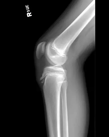
The main research method is x-ray of the hip joints. Pictures are taken in standard projections and Lauenstein's projection ("frog pose"). The X-ray picture for this disease depends on the severity of the pathological process and its stage.

Perthes disease on x-ray
A more informative diagnostic method at an early stage of Perthes disease is magnetic resonance imaging of the hip joint, which makes it possible to accurately assess the state of bone and soft tissues.
Treatment
Expectant tactics in Perthes disease is justified only in children under 6 years of age with minimal changes on radiographs and a mild clinical picture.
In all other cases, patients need long-term conservative therapy, which lasts several years (on average, 2.5-3 years). It includes:
- unloading the limb using plaster casts, skeletal traction;
- medication and non-medication methods to improve blood supply to the femoral head;
- maintaining muscle tone;
- stimulation of the process of resorption of necrotic tissue;
- stimulation of osteogenesis (the formation of new bone tissue).
In the course of conservative treatment of Perthes disease, methods of physiotherapy are actively used (exercise therapy, massage, ozokerite, mud therapy, electrophoresis with phosphorus and calcium, UHF).

In case of Perthes disease, therapeutic massage is actively used
Surgical treatment of Perthes disease is prescribed for children over 6 years old in the presence of chronic hip subluxation or severe deformity of the hip joint.
Possible complications and consequences
One of the most serious complications of Perthes disease is the development of deforming osteoarthritis of the hip joint (coxarthrosis), leading to gait disorder and the appearance of persistent pain.
Children with Perthes disease are prone to obesity, as they have to lead a sedentary lifestyle for a long time. Therefore, they are advised to adhere to a diet that is low in fat and carbohydrates.
Forecast
The prognosis depends on the location and size of the necrotic area. With minor necrosis and timely treatment, the hip joint is usually fully restored.
With severe aseptic necrosis, the femoral head disintegrates into several separate fragments. Subsequently, they grow together, giving the head an irregular shape, which causes an anatomical discrepancy between the femoral head and the acetabulum. This limits the support function of the leg, contributes to the development of contractures.
Prevention
There are no preventive measures to prevent the development of Perthes disease.
To prevent severe coxarthrosis, which is a complication of the underlying disease, patients are advised to limit physical activity on the hip joint throughout their lives. Patients with Perthes disease should not engage in running, jumping, or strenuous physical work, but exercise therapy and swimming are very useful for them. Regular spa treatment also helps maintain an acceptable level of health.
YouTube video related to the article:

Elena Minkina Doctor anesthesiologist-resuscitator About the author
Education: graduated from the Tashkent State Medical Institute, specializing in general medicine in 1991. Repeatedly passed refresher courses.
Work experience: anesthesiologist-resuscitator of the city maternity complex, resuscitator of the hemodialysis department.
The information is generalized and provided for informational purposes only. At the first sign of illness, see your doctor. Self-medication is hazardous to health!


