- Author Rachel Wainwright wainwright@abchealthonline.com.
- Public 2023-12-15 07:39.
- Last modified 2025-11-02 20:14.
Intracranial hematoma

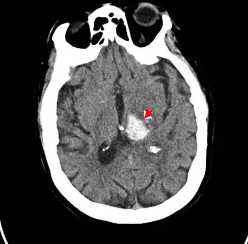
An intracranial hematoma (blood tumor) is an accumulation of blood in the skull cavity that reduces the intracranial space and contributes to compression of the brain. Such accumulations of blood occur as a result of rupture of an aneurysm, vascular trauma and hemorrhage - in a tumor, of an infectious origin or as a result of a stroke.
A feature of intracranial hematoma is that clinical manifestations do not appear immediately, but after a certain period of time.
The biggest danger of an intracranial hematoma is that it puts significant pressure on the brain. As a result, cerebral edema can form, with damage to the brain tissue and its subsequent destruction.
Types of intracranial hematomas
Hematomas are:
- acute - symptoms appear within 3 days from the moment of formation;
- subacute - symptoms appear for 21 days;
- chronic - the onset of symptoms occurs after 21 days from the moment of formation.
By size, small hematomas (up to 50 ml), medium (50-100 ml) and large (more than 100 ml) are distinguished.
At the site of localization, hematomas are divided into:
- epidurals located above the dura mater of the brain;
- subdural, with localization between the brain substance and its hard shell;
- intracerebral and intraventricular, the place of localization of which falls directly on the brain substance;
- intracranial hematomas of the brain stem;
- diapedetic hematomas resulting from hemorrhagic impregnation, while the integrity of the vessels is not disturbed.
The main causes of intracranial hematoma
The main cause of intracranial hematoma is disease or injury.
So, subdural hemorrhage often occurs as a result of rupture of veins connecting the brain and the venous system, as well as the sinuses of the dura mater. The result is a hematoma that compresses the brain tissue. Since blood from the vein accumulates slowly, the symptoms of subdural hematoma may not appear for several weeks.
An epidural hematoma usually results from a ruptured artery or vessel between the skull and the outer surface of the dura mater. The blood pressure in the arteries is higher than in the veins, so blood flows out of them faster. An epidural hematoma rapidly grows in size and increases pressure on the brain tissue. Symptoms usually appear fairly quickly, sometimes even within hours.
Intracerebral hematoma is formed as a result of penetration of blood into the brain. If a cerebral hemorrhage occurs as a result of injury, the white matter of the brain is predominantly affected. As a result of such damage, neurites are ruptured, which cease to transmit impulses to different parts of the body. Intracerebral hematoma can also form as a result of hemorrhagic stroke. In this case, hemorrhage occurs from an unevenly thinned artery wall and high pressure blood enters the brain tissue and fills the free space. Such a hematoma can form anywhere in the brain.
Thinning and rupture of blood vessels occur, as a rule, as a result of tumors, infections, angioedema, atherosclerotic lesions, etc.
Sometimes diapedesic hemorrhages may occur as a result of increased vascular permeability (with a change in the coagulation properties of blood or tissue hypoxia). This leads to the formation of accumulations of blood around the damaged vessels, which often merge, and an intracranial hematoma is formed.
Intracranial hematoma symptoms
Often the symptoms of intracranial hematoma appear after a certain period of time. The main symptoms depend on the nature of the intracranial hematoma and its size. Since the hematoma predominantly develops as a result of traumatic injury, then the symptoms generally predominate, characteristic of brain damage. In addition, the symptoms of hematoma may differ depending on the age of the patient.

With an epidural hematoma, symptoms appear quickly. Patients suffer from severe headache, drowsiness, confusion. Often, patients with epidural hematoma become comatose. When a hematoma forms more than 150 ml, a person dies. There is a progressive dilatation of the pupil on the side of the hematoma. The patient may experience epileptic seizures, paralysis and progressive paresis. In children, the symptoms of an epidural hematoma are as follows: there is no primary loss of consciousness, edema develops very quickly and requires immediate surgical treatment of an intracranial hematoma.
With the formation of a subdural hematoma, symptoms usually do not appear immediately, and the initial lesion seems insignificant. Symptoms usually begin to appear after a few weeks. In young children, an increase in the size of the head may be observed. In elderly patients, a subacute course of hematoma is observed. Young patients experience a headache, later vomiting and nausea, epileptic seizures and convulsions may appear. There may be an expansion of the pupil from the side of the injury, but not always. Small intracranial hematomas can resolve on their own, while large hematomas need to be emptied.
With intracerebral hematoma as a result of hemorrhagic stroke, the symptoms depend on the lesion focus. The most common symptoms are headache (predominantly on one side), wheezing, loss of consciousness, and paralysis, seizures and vomiting. If the brainstem is damaged, intracranial hematoma cannot be treated, and the patient dies.
With an intracranial hematoma that has formed as a result of extensive trauma, the symptoms are usually the following: headache, loss of consciousness, vomiting, nausea, epileptic seizures, convulsions. It is usually possible to determine the localization of such a hematoma only as a result of surgery.
When a hematoma forms due to a ruptured aneurysm, the main symptom is a sharp and sharp pain in the head (like a dagger strike).
Intracranial hematoma treatment
Mostly the treatment of intracranial hematoma involves surgery. The type of surgery often depends on the nature of the hematoma.
After surgery, your doctor will prescribe anticonvulsant drugs to prevent or control post-traumatic seizures. It happens that such seizures begin in a patient even a year after the injury. For a while, the patient may have amnesia, headache and impaired attention.
The recovery period after an intracranial hematoma is usually very long. In adult patients, the recovery period takes at least six months. Children tend to recover much faster.
YouTube video related to the article:
The information is generalized and provided for informational purposes only. At the first sign of illness, see your doctor. Self-medication is hazardous to health!


