- Author Rachel Wainwright wainwright@abchealthonline.com.
- Public 2023-12-15 07:39.
- Last modified 2025-11-02 20:14.
Arthrosis of the hip joint
The content of the article:
- Risk factors and causes of arthrosis of the hip joint
- Forms of the disease
- Stages
- Symptoms of arthrosis of the hip joint
- Diagnostics
- Treatment of arthrosis of the hip joint
- Potential consequences and complications
- Forecast
- Prevention
Arthrosis of the hip joint (deforming arthrosis, coxarthrosis, osteoarthritis) is a slowly progressive degenerative-dystrophic disease, leading over time to destruction of the affected joint, persistent pain and limited mobility.
The disease affects people over 40, women get sick several times more often than men.
In the general structure of arthrosis, arthrosis of the hip joint plays a leading role. This is due to the widespread congenital pathology of the hip joints (dysplasia), as well as the significant physical exertion to which these joints are subject.

Arthrosis of the hip joint is the most common disease of the musculoskeletal system
Risk factors and causes of arthrosis of the hip joint
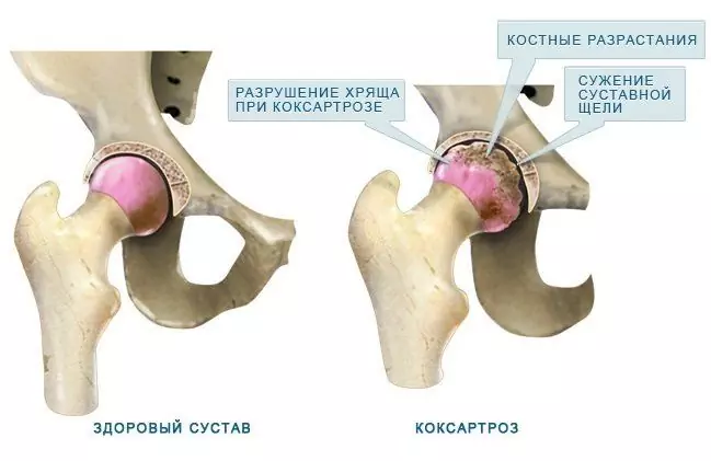
In the pathological mechanism of the development of arthrosis of the hip joint, the main role belongs to a change in the physicochemical characteristics of the synovial (intra-articular) fluid, as a result of which it becomes thicker and more viscous. This impairs its lubricating properties. When moving, the articular cartilage surfaces begin to rub against each other, become rough, and become covered with cracks. Small particles of hyaline cartilage break off and enter the articular cavity, causing the development of aseptic (non-infectious) inflammation in it. As the disease progresses, bone tissue is drawn into the inflammatory process, which leads to aseptic necrosis of areas of the femoral head and the surface of the acetabulum, the formation of osteophytes (bone growths), which intensify inflammation and cause severe pain during movement.
At a late degree of arthrosis of the hip joint, the inflammation spreads to the surrounding tissue of the joint (vessels, nerves, ligaments, muscles), which leads to the appearance of signs of periarthritis. As a result, the hip joint is completely destroyed, its functions are lost, movement in it stops. This condition is called ankylosis.
Causes of arthrosis of the hip joint:
- congenital dislocation of the hip;
- dysplasia of the hip joint;
- aseptic necrosis of the femoral head;
- Peters disease;
- hip joint injuries;
- infectious arthritis of the hip joint;
- gonarthrosis (deforming osteoarthritis of the knee joint);
- osteochondrosis;
- excess weight;
- professional sports;
- flat feet;
- rachiocampsis;
- sedentary lifestyle.
Pathology is not inherited, but the child inherits from his parents the structural features of the musculoskeletal system, which can cause arthrosis of the hip joint under conditions conducive to this. This explains the fact of the existence of families, the incidence of which is higher than in the general population.
Forms of the disease
Depending on the etiology, arthrosis of the hip joint is divided into primary and secondary. Secondary arthrosis develops against the background of other diseases of the hip joint or its injuries. The primary form is not associated with the previous pathology, the cause of its development is often not established, in this case they speak of idiopathic arthrosis.
Coxarthrosis is unilateral or bilateral.
Stages
During arthrosis of the hip joint, there are three stages (degrees):
- Initial - pathological changes are not very pronounced, provided timely and adequate treatment they are reversible.
- Progressive coxarthrosis is characterized by a gradual increase in symptoms (pain in the joint and impaired mobility), changes in the joint tissues are already irreversible, but therapy can slow down degenerative processes.
- Final - movement in the joint is lost, ankylosis is formed. Treatment is possible only by surgery (replacement of the joint with an artificial one).

Arthrosis of the hip joint is divided into 3 degrees
Symptoms of arthrosis of the hip joint
The main signs of arthrosis of the hip joint:
- pain in the groin, hip and knee;
- feeling of stiffness in the affected joint and limitation of its mobility;
- lameness;
- restriction of abduction;
- atrophic changes in the muscles of the thigh.
The presence of certain symptoms of arthrosis of the hip joint, as well as their severity, depend on the degree of the disease.

Pain in the groin and hip area may indicate arthrosis of the hip joint
At the 1st degree of arthrosis of the hip joint, patients complain of pain arising under the influence of physical activity (long walking, running) in the affected joint. In some cases, the pain is localized to the knee or thigh. After a short rest, the pain goes away on its own. The range of motion of the limb is completely preserved, the gait is not disturbed. The radiograph shows the following changes:
- a slight uneven decrease in the lumen of the joint space;
- osteophytes located along the inner edge of the acetabulum.
No changes in the neck and head of the femur were detected.
With the II degree of arthrosis of the hip joint, pain appears at rest, including at night. After physical exertion, the patient begins to limp, a characteristic "duck" gait is formed. The so-called starting pains appear - after a long period of immobility, the first few steps cause pain and discomfort, which then pass, and then return after a prolonged load. The range of motion is limited in the affected joint (abduction, internal rotation). The radiograph shows that the joint space is unevenly narrowed and its lumen is 50% of the norm. Osteophytes are located both along the inner and outer edges of the glenoid cavity, going beyond the borders of the cartilaginous lip. The contours of the femoral head become uneven due to deformation.
With III degree of arthrosis of the hip joint, the pain is intense and constant, which does not stop at night. Walking is significantly difficult, the patient is forced to lean on a cane. The range of motion in the affected joint is sharply limited, later completely stops. Due to the atrophy of the thigh muscles, the pelvis deviates in the frontal plane and the limb is shortened. Trying to compensate for this shortening, when walking, patients are forced to deflect the trunk towards the lesion, which further increases the load on the diseased joint. Radiographs show multiple bony growths, a significant narrowing of the joint space and a pronounced increase in the femoral head.
Diagnostics
Diagnosis of arthrosis of the hip joint is based on the data of the clinical picture of the disease, the results of medical examination and instrumental studies, among which the main importance belongs to imaging methods - radiography, computed or magnetic resonance imaging. They allow not only to determine the presence of arthrosis of the hip joint and assess its degree, but also to identify the possible cause of the disease (trauma, juvenile epiphysiolysis, Peters disease).
Differential diagnosis of arthrosis of the hip joint with other diseases of the musculoskeletal system is rather difficult. At II and III degrees of arthrosis of the hip joint, muscle atrophy develops, which can cause intense pain in the knee joint, characteristic of gonitis or gonarthrosis (diseases of the knee joint). For the differential diagnosis of these conditions, palpation of the knee and hip joints is performed, the volume of movement in them is determined, and they are also examined radiographically.

Arthrosis of the hip joint on x-ray
In diseases of the spine, in some cases, compression of the nerve roots of the spinal cord occurs with the development of pain syndrome. Pain can radiate to the area of the hip joint and mimic the clinical picture of its lesion. However, the nature of pain in radicular syndrome is somewhat different than in arthrosis of the hip joint:
- pain occurs as a result of lifting weights or a sharp awkward movement, and not under the influence of physical exertion;
- the pain is localized in the gluteal, not the groin area.
With radicular syndrome, the patient can safely move his leg to the side, while with arthrosis of the hip joint, abduction is limited. A characteristic sign of radicular syndrome is a positive symptom of tension - the appearance of sharp pain when a patient is lying on his back to raise a straight leg.
Arthrosis of the hip joint should be differentiated with trochanteric bursitis (trochanteritis). Trochanteric bursitis develops faster, over several weeks. It is usually preceded by significant physical activity or injury. The pain with this disease is much more pronounced than with arthrosis of the hip joint. At the same time, limb shortening and limitation of its mobility are not detected.
The clinical picture of atypical reactive arthritis and ankylosing spondylitis may resemble the clinical manifestations of arthrosis of the hip joint. However, pain occurs in patients mainly at night or at rest, while walking does not increase, but, on the contrary, decreases. In the morning, patients notice stiffness in the joints, which disappears after a few hours.
Treatment of arthrosis of the hip joint
Orthopedists are involved in the treatment of arthrosis of the hip joints. With I and II degrees of the disease, conservative therapy is indicated. With severe pain syndrome, patients are prescribed non-steroidal anti-inflammatory drugs in a short course. They should not be taken for a long time, since they are not only capable of having a negative effect on the organs of the gastrointestinal tract, but also suppress the regenerative abilities of hyaline cartilage.
The treatment regimen for arthrosis of the hip joint includes chondroprotectors and vasodilators, which create optimal opportunities for the restoration of damaged cartilage tissues. With severe muscle spasm, central muscle relaxants may be required.

Chondroprotectors are prescribed for the treatment of arthrosis of the hip joint
In cases where it is not possible to stop the pain syndrome with non-steroidal anti-inflammatory drugs, they resort to intra-articular injections of corticosteroids.
Local treatment of arthrosis of the hip joint using warming ointments can reduce muscle spasm and somewhat relieve pain due to a distracting effect.
In the complex therapy of arthrosis of the hip joint, physiotherapeutic methods are also used:
- magnetotherapy;
- inductothermy;
- UHF;
- laser therapy;
- ultrasound treatment;
- massage;
- physiotherapy;
- manual therapy.
Diet food for arthrosis of the hip joint is aimed at correcting body weight and normalizing metabolic processes. Weight loss reduces the stress on the hip joints and thus slows the progression of the disease.
To relieve stress on the affected joint, the doctor may recommend that patients walk with support on crutches or a cane.
With III degree of arthrosis of the hip joint, conservative treatment is ineffective. In this case, improving the patient's condition, returning him to normal mobility is possible only as a result of surgical intervention - replacing the destroyed joint with an artificial one (joint arthroplasty).

For grade 3 arthrosis, joint replacement is indicated
Potential consequences and complications
The most serious complication of progressive arthrosis of the hip joint is disability due to loss of motion in the joint. With bilateral coxarthrosis, the patient loses the ability to move independently and needs constant outside care. Prolonged stay in bed in one position creates the prerequisites for the occurrence of congestive (hypostatic) pneumonia, which is difficult to treat and can lead to death.
Forecast
Arthrosis of the hip joints is a progressive chronic disease that can be completely cured only in the early stages, provided the cause of the disease is eliminated. In other cases, therapy can slow down its course, however, over time, it becomes necessary to implant hip joint endoprostheses. Such operations in 95% of cases provide complete restoration of limb mobility, restore the patient's working capacity. The service life of modern prostheses is 15-20 years, after which they must be replaced.
Prevention
Prevention of arthrosis of the hip joint is aimed at eliminating the causes that can lead to the development of this disease, and includes:
- timely detection and treatment of diseases and injuries of the hip joint;
- rejection of a sedentary lifestyle, regular, but not excessive physical activity;
- control of body weight;
- balanced diet;
- rejection of bad habits.
YouTube video related to the article:

Elena Minkina Doctor anesthesiologist-resuscitator About the author
Education: graduated from the Tashkent State Medical Institute, specializing in general medicine in 1991. Repeatedly passed refresher courses.
Work experience: anesthesiologist-resuscitator of the city maternity complex, resuscitator of the hemodialysis department.
The information is generalized and provided for informational purposes only. At the first sign of illness, see your doctor. Self-medication is hazardous to health!


