- Author Rachel Wainwright wainwright@abchealthonline.com.
- Public 2023-12-15 07:39.
- Last modified 2025-11-02 20:14.
Eye pressure: symptoms of increase and decrease, causes, normalization
The content of the article:
- Increasing and decreasing pressure in the eyes
- Measurement of intraocular pressure
- Signs of an increase and decrease in intraocular pressure
- Treatment of high and low eye pressure
- Video
Eye pressure - the pressure of the fluid in the eye (synonym - intraocular pressure). This indicator is of no small importance for people who are at risk for glaucoma, since high intraocular pressure is one of the main risk factors for the development of this disease.
The level of intraocular pressure depends on the amount of fluid consumed, the drugs taken, the presence of bad habits (in particular, alcohol abuse, the use of marijuana leads to an increase).

To measure intraocular pressure, you need to see an ophthalmologist
Normal intraocular pressure is 10-20 mm Hg. Art. Physiologically, it increases slightly with age. In addition, the indicator changes throughout the day, but the difference usually does not exceed 3 mm Hg. Art. A slight increase in intraocular pressure is observed in the morning, by the middle of the day the indicator decreases, the lowest intraocular pressure is observed at night.
Increasing and decreasing pressure in the eyes
An increase in intraocular pressure is called ocular hypertension (hypertension). What is ocular hypertension and why is this condition? It can develop under the influence of a genetic predisposition, inflammatory eye diseases, and taking certain medications. The reasons for the development of ocular hypertension include increased blood pressure, prolonged work at the computer, prolonged viewing of television programs, increased intracranial pressure, exposure to stress, physical overstrain, disorders of the cardiovascular, central nervous, urinary systems, hypothyroidism, intoxication of the body, menopause.
Typically, ocular hypertension is recorded in adult patients (usually in the age group over 40).
Decreased intraocular pressure (ocular hypotension or hypotension) is a rather rare pathological condition, usually associated with the patient's blood pressure level. Intraocular pressure can decrease with fluid leakage and phthisis of the eyeball, a decrease in pressure in the capillaries of the eye. Risk factors are inflammation in the eyeball, surgery, injury to the eyeball, foreign body ingestion, eye development abnormalities, retinal detachment, diseases of the urinary system, dehydration of the body.
Measurement of intraocular pressure
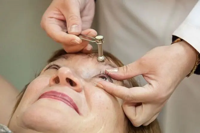
For the primary determination of increased intraocular pressure, palpation of the eyeball can be performed through the eyelids. The indicator is accurately measured in two ways:
- ocular pressure tonometry according to Maklakov;
- non-contact tonometry (air jet tonometry) - usually used when glaucoma is suspected.
It should be borne in mind that the eye pressure in the right and left eyes may differ, therefore the indicator is determined in both eyes. The measurement of intraocular pressure is influenced by the stiffness and thickness of the cornea. For this reason, some types of manipulations (for example, photorefractive keratectomy) can lead to distortion of the measurement results of this indicator, which must also be borne in mind when diagnosing.
Signs of an increase and decrease in intraocular pressure
Hypertension of the eye is divided into several forms:
- stable - intraocular pressure constantly goes beyond the upper limit of the norm, which may indicate glaucoma;
- labile - jumps in intraocular pressure are characteristic, when the indicator alternately rises, then returns to normal;
- transient - a short-term increase in intraocular pressure.
Pathology often does not manifest itself for a long time. With a significantly increased intraocular pressure, the patient has a reduction in the field of vision, impaired twilight vision, pain in the temples and / or eyebrows, rainbow arches in front of the eyes when looking at light. In addition, patients with ocular hypertension are worried about severity, distention, burning in the eyes, and discomfort even with insignificant visual stress. With the progression of the pathological process, the patient develops an intense headache resembling a migraine. There is often severe pain in one or both eyes, and redness.
How reduced intraocular pressure manifests itself depends on the cause of its development. Dry eyes are common when an infection is present. With severe hypotension, the eyeball sinks. With a prolonged gradual decrease in intraocular pressure, any manifestations may be absent altogether. In some patients, only visual impairment indicates ocular hypotension.
With the frequent appearance of one or another suspicious symptom of eye pressure, you should not guess what he is talking about, but you should contact an ophthalmologist and undergo an examination.
Treatment of high and low eye pressure
What to do in case of increased intraocular pressure? It is not recommended to treat the pathology on your own, since the consequences can be the most unfavorable, including complete loss of vision in one eye or both.
To normalize high ocular pressure, they resort to drug therapy, in the appointment of which the cause of the pathology is taken into account. They usually start with the use of eye drops.
The outflow of fluid is facilitated by prostaglandins. The effect of their use occurs within two hours. Possible side effects include eye redness and discoloration of the iris.
Also, with increased intraocular pressure, cholinomimetics are prescribed. Side effects of their use can be pain in the temples and superciliary arches, narrowing of the visual field.
In some cases, beta-blockers are shown, the effect of which is manifested much faster, however, the patient may develop disorders of the respiratory and cardiovascular system, which must be taken into account when treating patients with concomitant pathology.
A reduction in the production of ocular fluid can be achieved with carbonic anhydrase inhibitors. Drugs in this group are not prescribed for pathologies of the urinary system, as they can worsen its functions.
Usually, patients with increased intraocular pressure are shown taking vitamin complexes.
If the visual functions are preserved, exercises for the eyes are usually prescribed, the patient is advised to avoid traumatic sports, activities that require eye strain; it is necessary to reduce the time of watching TV and working at the computer, wearing glasses with protective properties. Patients should spend more time in the fresh air, eat right, lead a moderately active lifestyle.
In case of serious damage to the visual analyzer, the patient may be shown surgical intervention - stretching the trabecula with a laser, laser excision of the iris.
It is possible to normalize slightly increased intraocular pressure at home with the help of folk remedies. For these purposes, an infusion of meadow clover (taken daily at night), tincture of golden mustache (1 dessert spoon before meals), kefir with the addition of cinnamon are used. It should be remembered that any methods of traditional medicine can only be used after consulting your doctor.

A persistent increase in intraocular pressure may indicate the development of glaucoma.
With reduced eye pressure, depending on the cause, antibacterial drugs, medicines to strengthen blood vessels, stimulants that increase intraocular pressure, vitamins, etc. may be prescribed. Pathology caused by trauma may require surgical intervention.
People with atherosclerotic vascular lesions and hyperopia (farsightedness) should be especially attentive to the state of the visual analyzer, this combination is characteristic of elderly people. In order to prevent relapses, it is necessary to avoid excessive physical and mental stress, limit the use of fatty, fried, salty and smoked foods, stop smoking, drinking strong tea, and alcoholic beverages. It is recommended to drink 1.5 liters of clean water per day.
To prevent the development of intraocular pressure disorders, you need to take five-minute breaks every hour of working at the computer, during which they do eye exercises and / or light eyelid massage. The diet should regularly include carrots, blueberries, fish. Patients from the risk group are recommended to undergo an ophthalmological examination once a year, including a study of visual acuity, visual fields, examination of the fundus.
Video
We offer for viewing a video on the topic of the article.

Anna Aksenova Medical journalist About the author
Education: 2004-2007 "First Kiev Medical College" specialty "Laboratory Diagnostics".
Found a mistake in the text? Select it and press Ctrl + Enter.


