- Author Rachel Wainwright wainwright@abchealthonline.com.
- Public 2024-01-15 19:51.
- Last modified 2025-11-02 20:14.
Chickenpox scars
The content of the article:
- What type are scars after chickenpox
- Scar formation
- External manifestations
-
Removal methods
- Medication methods of treatment
- Physiotherapy treatments for scars
- Surgical removal
- Video
Chickenpox scars are one of the complications. Chickenpox is an acute highly contagious infectious disease caused by a virus of the Herpesviridae family (varicella zoster). The causative agent is able to penetrate into the growth layer of the epidermis with the formation of papulovesicular rash. As the vesicles heal, they become covered with crusts, under which normal tissue healing occurs. Most chickenpox leaves no visible traces behind. However, in the case of tearing off the crust due to intense itching, a deep ulcer is formed at the site of the vesicle, which, during the healing process, will be replaced not by a typical stratum corneum, but by connective tissue - this is how scars or scars appear.

Chickenpox scars usually look like pits in the skin (atrophic scar)
What type are scars after chickenpox
All scars are divided into two main types - physiological (normotrophic) and pathological. Pathological, in turn, are of three subspecies:
- atrophic;
- hypertrophic;
- keloid.
Classic chickenpox scars are atrophic. Other types of scars in this disease occur only with the addition of a secondary infection and a complicated course of chickenpox (pustular, gangrenous or bullous form).
Scar formation
The 3 main stages of scar formation in the healing of any wound:
| Phase | Characteristic |
| Exudation and inflammation |
The blood clotting system is activated, as the superficial vasculature, clot formation and the creation of a matrix of collagen for further wound healing are often affected. After some time, the formed clot is destroyed (fibrinolysis), this process is associated with the release of numerous growth factors (transforming growth factor β, epidermal growth factor). |
| Proliferation | Formation of young connective tissue at the site of injury, which at this stage is represented mainly by type III collagen, which has good extensibility and elasticity. |
| Reorganization phase | Typically, after 3-6 months, the tissue matures and type III collagen is replaced by less elastic type I collagen. This stage is associated with the formation of a final scar at the site of the former damage to the skin. |
External manifestations
There are several options for atrophic scars, depending on the shape:
- chipped;
- rectangular;
- rounded;
- striae.
They are characterized by atrophy of the dermis and severe fibrosis (there are no cells and vessels at the site of injury). The external manifestations of atrophic scars (see photo) are as follows:
- Scars are located below the surface of the skin, that is, they form depressions or, as they are often called, pits.
- Diameter from 2 mm to 1 cm.
- Do not differ in color from the surrounding skin.
- They tend to merge.
- Localization depends on the initial smallpox, any part of the body can be affected.
Hypertrophic and keloid scars are more noticeable, they protrude significantly above the skin surface, their color is variable (depigmented, hyperpigmented or reddish when vascular tissue grows). The probability of their occurrence after chickenpox is less than 1%.
Removal methods
There are three ways to remove chickenpox scars on your face or other body parts:
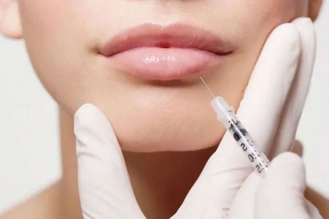
- conservative (gel, ointment, patch, injections);
- physiotherapy;
- surgical.
Medication methods of treatment
| Facilities | Act | Examples of |
| Corticosteroid drugs | They are mainly used for patients with keloid scars, but are acceptable for the treatment of other forms. They have a pronounced anti-inflammatory effect, regulate metabolic processes in tissues. |
1. Hydrocortisone - used for local injections. 2. Triamcinolone acetate - point injections into the damaged area with an interval of 4-6 weeks. |
| Enzyme preparations | The action of the drugs is aimed at restoring the water balance in tissues, improving its trophism and increasing the elasticity of scar-altered areas. Suitable for all types of pathological scars. | Glycosaminoglycans, collagenases, hyaluronidases (Longidase, Imoferase). |
| Immunomodulators | Suitable for all types of pathological scars. They inhibit the production of collagen (I and III types). | Interferon-α2b is injected into a pathological scar after its excision. |
| Vitamin therapy | Used for all types of pathological scars. They have a pronounced wound-healing effect, inhibit the proliferation of fibroblasts, and stimulate the proliferation of normal skin cells. | Retinol by intradermal injection. |
| Flavonoid compounds | They are able to somewhat inhibit collagen production and stabilize cell membranes. | Plant-based compounds: Quercetin, Protocatechin. |
| Combined drugs | They are able to influence the synthesis of collagen and, thus, prevent the formation of coarse keloid scars, some are capable of partially destroying the connective tissue. |
1. Contractubex - contains onion extract, heparin, allantoin. It is required to process the site at least 2 times a day. 2. Fermenkol. It is an enzyme complex obtained from the digestive organs of the Kamchatka crab. Smear the affected area at least 2 times a day. 3. Dermatics. The preparation is based on organosilicon compounds. |

Very rarely, after chickenpox, hypertrophic scars can form
Physiotherapy treatments for scars
| Method | Description |
| Cryodestruction | Exposure to the pathological focus with liquid nitrogen and the formation of microcrystals inside the cell as a result, which causes damage to the vascular bed, cell death. Most often used in combination with corticosteroid injections. After the first procedure, the defect is reduced by only 50%. |
| Laser therapy | The optimal method of correction for all types of pathological scars. For the treatment of atrophic scars, ablative (carbon dioxide, erbium) and non-ablative lasers (Q-switched neodymium YAG laser 1064 nm, diode 1450 nm, neodymium 1320 nm) are often used. On average, a course of 3 to 5 procedures is prescribed. An equally good result is obtained with the use of a CO2 laser and plasmolifting (introduction of platelet-rich plasma into the site of injury, which causes enhanced tissue regeneration). In this case, tissue restructuring and type I collagen synthesis occur. |
| Cosmetological methods using concentrated acids and alkalis (peeling, mesotherapy, dermabrasion) | They are used for lesions of small diameter, it is permissible to start their use at the 3rd phase of scar formation, which means that they are not able to influence the process of formation of colloidal tissue itself. The methods are less effective than laser exposure. The use of chemical peels is associated with the risk of chemical burns if the technique is not followed, so it is advisable to avoid them. |
| Bucky therapy | Ultra-soft X-rays (the so-called boundary rays), the energy range of which is 5-15 keV. It is more often used for keloid and hypertrophic scars, since the method allows them to level with the skin surface and return to normal color. After the first session, an inflammatory reaction is possible, which independently regresses within 7-10 days. |
| Fractional photothermolysis | The type of laser exposure is currently the most modern method of scar correction. Under the influence of rays of a certain wave on 1 cm2 of the skin, about 2000 microtraumatic zones with a diameter of 70-100 microns each are formed. This starts the process of collagen reorganization, the scar is smoothed with an almost complete return of the skin to its original state. To get rid of a scar, an average of 2-3 procedures are required. |

A good cosmetic effect is obtained after laser removal of scars
Surgical removal
Surgical treatment in the case of atrophic scars is rarely resorted to. Why? The reason is that surgery itself leaves scars. This option is often used in the case of single keloid or hypertrophic scars in adult patients, when other methods are ineffective. In this case, surgical intervention will completely radically get rid of the formation.
Video
We offer for viewing a video on the topic of the article.

Anna Kozlova Medical journalist About the author
Education: Rostov State Medical University, specialty "General Medicine".
Found a mistake in the text? Select it and press Ctrl + Enter.


