- Author Rachel Wainwright [email protected].
- Public 2023-12-15 07:39.
- Last modified 2025-11-02 20:14.
Echocardiography

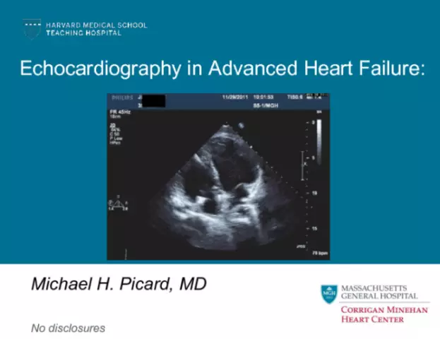
Echocardiography is one of the methods of ultrasound diagnostics used to study functional and morphological changes in the heart and its structures. This diagnostic method allows you to assess the condition of the heart valves in imaging mode. Echocardiogram is widely used for early diagnosis of cardiac diseases, which makes it possible to identify pathology in time.
Indications for cardiac echocardiography
An ultrasound examination of the heart is prescribed for:
- Murmurs of varying degrees and localization in the heart;
- Feverish conditions of uncertain cause;
- Detection of changes in the ECG;
- Suspected congenital or acquired heart defects;
- Visible abnormalities on the roentgenogram - with an increase in the size of the heart or its structures, a modified aorta, an accumulation of calcifications;
- Arrhythmias, including felt arrhythmias;
- Hereditary serious pathologies of the cardiovascular system;
- Complaints of fainting, chest pain, leg swelling, severe shortness of breath and frequent dizziness;
- Increased blood pressure;
- Postponed myocardial infarction;
- Suspicion of a heart tumor, as well as enlargement of the thoracic aorta.

Echocardiography of the heart can detect the presence of neoplasms and diagnose various lesions. In addition, an echocardiogram is used to monitor the condition in ischemic disease, congenital heart defects, after a heart attack, with cardiomyopathy and arterial hypertension.
Also, cardiac echocardiography is recommended for regular sports training and other activities that cause increased stress on the heart or with constant emotional stress. In childhood, in the process of intensive development and growth in a child, and especially if there is a suspicion of congenital heart disease, an echocardiogram is also prescribed.
Fetal echocardiography
Congenital heart defects, which are one of the leading causes of perinatal mortality, can be diagnosed by fetal echocardiography. This procedure is harmless to the fetus and makes it possible to assess intracardiac hemodynamics in the prenatal period, as well as to dynamically monitor the indicators during pregnancy when systolic heart murmurs are detected. This method of echocardiogram of the heart and blood vessels is often used to assess the state of organs before the birth of a child, which allows a timely diagnosis and treatment to begin almost immediately after birth. Echocardiography of the fetus examines the heart and blood vessels in detail, revealing congenital disorders in its work.
If necessary, fetal echocardiography is performed at 18 to 22 weeks of gestation. The indications for the appointment of diagnostics are:
- Congenital heart defects in relatives;
- Miscarriages before the current pregnancy;
- Diabetes;
- Taking antibiotics, as well as medicines for epilepsy in the first trimester;
- Deviations recorded on the planned ultrasound of the fetus at 20 weeks.
Fetal echocardiography is interpreted by cardiologists or a geneticist.
Methods for conducting echocardiography of the heart
Depending on the indications, cardiac echocardiography can be performed using several methods that allow assessing various parameters:
- Echocardiography in M-mode - the size of the heart and the systolic work of the ventricles;
- Two-dimensional echocardiography - wall thickness and dimensions of the heart cavities, ventricular contractility, valve condition, and the presence or absence of cavity thrombosis;
- Transesophageal - the back wall of the heart, usually after a heart attack of the lower heart wall;
- Intravascular ultrasound - the state of the coronary vessels;
- Volumetric modeling of the heart;
- Doppler research - central dynamics;
- Stress - changes in various segments of the heart caused by physical activity;
- Contrast echocardiography - the state of the heart chambers.
Echocardiography is interpreted by a cardiologist. Based on the diagnostics performed, a researcher can have more than one hundred initial and calculated indicators and objectively evaluate:
- Contractility;
- Valve condition and function;
- The functional state of the myocardium;
- The presence of intracardiac blood clots;
- Indicators of pumping function and left ventricular contractility in dynamics;
- The dimensions of the cavities of the heart;
- The thickness of the walls of the heart;
- The presence of scars;
- The degree of hypertrophy of the cardiac chambers.
Contraindications to echocardiography
Echocardiography is a safe diagnostic procedure with no absolute contraindications. However, if the chest deformity, which interferes with normal diagnosis, as well as some allergic and inflammatory skin lesions in the heart, it can be difficult to carry out.
Found a mistake in the text? Select it and press Ctrl + Enter.


