- Author Rachel Wainwright wainwright@abchealthonline.com.
- Public 2023-12-15 07:39.
- Last modified 2025-11-02 20:14.
Renal artery
The renal artery is a paired terminal blood vessel that extends from the lateral surfaces of the abdominal aorta and supplies blood to the kidney. The renal arteries carry blood to the apical, posterior, inferior, and anterior segments of the kidney. Only 10% of the blood goes to the medulla of the kidney, and most (90%) to the cortex.

Renal artery structure
There are right and left renal arteries, each of which is divided into posterior and anterior branches, and these in turn are divided into segmental branches.
Segmental branches branch into interlobar branches, which break up into a vascular network, consisting of arched arteries. Interlobular and cortical arteries, as well as medullary branches, from which blood flows to the lobes (pyramids) of the kidney, depart from the arched arteries to the renal capsule. All together they form arcs from which the bringing vessels depart. Each bringing vessel branches into a tangle of capillaries, enclosed by the capsule of the glomerulus and the base of the renal tubule.
The outflowing artery also splits into capillaries. Capillaries entwine the kidney tubules, and then pass into the veins.
The right artery from the aorta runs forward and straight, and then goes to the kidney, obliquely and down, behind the inferior vena cava. The path of the left artery to the hilum is much shorter. It moves horizontally and from behind the left renal vein flows into the left kidney.
Renal artery stenosis
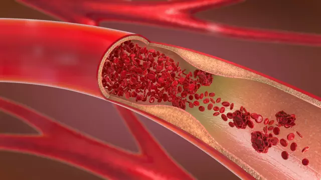
Partial occlusion of an artery or its main branches is called stenosis. Stenosis develops as a result of inflammation or compression of the artery by a tumor, dysplasia or atherosclerotic vasoconstriction. Fibromuscular dysplasias are a group of lesions in which there is a thickening of the middle, inner or subadential membranes of the vessel.
With stenosis of the renal arteries, the function of the kidney is disrupted due to its inadequate supply of blood. Kidney dysfunction often leads to the development of renal failure. Renal artery stenosis sometimes manifests itself as a sharp increase in blood pressure. But most often this disease is asymptomatic. Prolonged arterial stenosis can lead to azotemia. Azotemia manifests itself in confusion, weakness, and fatigue.
The presence of stenosis is usually determined using CT angiography, Doppler ultrasonography, urophragia, arteriography. Additionally, to identify the causes of the disease, urine analysis, biochemical and general blood tests are performed, and the concentration of electrolytes is determined.
To reduce pressure in stenosis, a combination of antihypertensive drugs with diuretics is usually prescribed. When the lumen of the vessel is narrowed by more than 75%, surgical methods of treatment are used - balloon angioplasty, stenting.
Renal artery denervation
To achieve a persistent antihypertensive effect, endovascular surgeons use the method of catheter sympathetic denervation of the renal arteries.
Renal artery denervation is an effective bloodless treatment for resistant hypertension. During the procedure, a catheter is inserted into the patient's femoral artery, which penetrates the arteries. Then, under short-term anesthesia, radiofrequency cauterization of the mouths of the arteries is performed from the inside. Cauterization destroys the connection of the afferent and efferent sympathetic nerves of the arteries with the nervous system, which leads to a weakening of the effect of the kidneys on blood pressure indicators. After cauterization, the conductor is removed, and the puncture site of the femoral artery is closed with a special device.
After denervation, there is a stable decrease in blood pressure by 30-40 mm Hg. Art. throughout the year.
Renal artery thrombosis
Renal artery thrombosis is the blockage of renal blood flow by a thrombus that has detached from extrarenal vessels. Thrombosis occurs with inflammation, atherosclerosis, trauma. In 20-30% of cases, thrombosis is bilateral.
With renal artery thrombosis, acute and severe pain occurs in the lower back, kidney, back, which spreads to the abdomen and side.
In addition, thrombosis can cause a sudden, significant increase in blood pressure. Very often, with thrombosis, nausea, vomiting, constipation appear, and body temperature rises.
Treatment of thrombosis is complex: anticoagulant treatment and symptomatic therapy, surgical intervention.
Renal artery aneurysm
A renal artery aneurysm is a saccular expansion of the lumen of a vessel due to the presence of elastic fibers in its wall and the absence of muscle fibers. Aneurysm is most often unilateral. It can be placed both intrarenally and extrarenally. Clinically, this pathology can be manifested by vascular thromboembolism and arterial hypertension.
For renal artery aneurysm, surgery is indicated. There are 3 types of surgery for this type of anomaly:
- artery resection;
- excision of the aneurysm with the replacement of its defect with a patch;
- aneurysmography - closure of the arterial wall with the tissues of the aneurysm left after preliminary excision of its main part
Aneurysmography is used for multiple vascular lesions and large aneurysms.
Found a mistake in the text? Select it and press Ctrl + Enter.


