- Author Rachel Wainwright wainwright@abchealthonline.com.
- Public 2023-12-15 07:39.
- Last modified 2025-11-02 20:14.
Inferior vena cava
The inferior vena cava is a wide vessel formed by the fusion of the left and right iliac veins at the level of the fourth to fifth vertebrae of the lumbar spine. The diameter of the inferior vena cava varies from 20 to 34 mm. The length of the chest part is 2-4 cm, the abdominal part is 17-18 cm.

The structure of the inferior vena cava
The vein is placed in the retroperitoneal space, behind the internal organs, to the right of the aorta. It passes behind the upper part of the duodenum, behind the mesentery root and the head (apex) of the pancreas and enters the hepatic furrow, absorbing the veins of the liver.
Passing through the hole of the same name in the tendon region of the diaphragm, the vein flows into the posterior region of the chest cavity. In this case, elastic, collagen and muscle fibers of the vein wall are woven into the wall of the diaphragm.
Having reached the pericardial cavity, the vein enters the right atrium. At the site of the entrance to the right atrium, the vena cava is slightly thickened. This vein has no valves.
The diameter of the inferior vena cava changes during the respiratory cycle. When you exhale, the vein expands, and when you inhale, it contracts. Changing the diameter of the inferior vena cava makes it easier to recognize and differentiate from other large veins.
Inferior vena cava system
The inferior vena cava system belongs to the most powerful system in the human body. It accounts for about 70% of the total venous blood flow.
The system of the inferior vena cava is formed by the vessels that collect blood from the abdominal cavity, the walls and organs of the pelvis, and the lower extremities.
This vein has parietal (parietal) and visceral (visceral) tributaries.
Partial tributaries include:
- lumbar veins (three to four on each side) - collect blood from the muscles and skin of the back, from the walls of the abdomen, as well as from the area of the vertebral plexus;
- phrenic veins - originate from the lower surface of the diaphragm;
- ilio-lumbar, lateral sacral, lower and upper gluteal veins - collect blood from the muscles of the abdomen, thigh and pelvis.
Visceral tributaries include:
- gonadal veins - ovarian and testicular veins that collect blood from the ovary (testicle);
- renal veins - connected at the level of the cartilage with the inferior vena cava between the lumbar vertebrae (first and second). The left renal vein is much longer than the right renal vein. She crosses the aorta in front.
- adrenal veins - the right vein enters the inferior vena cava, and the left vein connects to the renal vein.
- hepatic veins - carry blood from the liver.
All veins (except the largest ones) form numerous plexuses inside and outside the organs for the redistribution of blood. In case of damage to any vein, the blood flow is directed along collaterals (bypass paths).
Inferior vena cava thrombosis
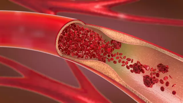
Thrombosis of the inferior vena cava accounts for about 11% of the total number of thrombosis of the veins of the pelvis and lower extremities. Vein thrombosis can be primary and secondary (depending on the cause of development).
Primary thrombosis develops as a result of a malignant or benign tumor, birth defects, vein trauma. The causes of secondary thrombosis can be the proliferation of a vein by a tumor or its compression. Often, secondary thrombosis of the inferior vena cava spreads ascending from other veins (smaller).
In medicine, thrombosis of the distal vein, as well as the renal and hepatic sections, is isolated. Thrombosis of the distal vein is manifested in cyanosis and edema of the lower extremities, lower abdomen, and lumbar region. Sometimes the swelling extends to the beginning of the chest. The upper limit of cyanosis and skin edema depends on the extent of the thrombosis.
With thrombosis of the renal segment of the vein, severe general disorders occur that can lead to death.
The development of thrombosis of the hepatic segment of the vein is most often accompanied by a violation of the basic functions of the liver and subsequent thrombosis of the portal vein. Symptoms of hepatic thrombosis include abdominal pain, enlargement of the spleen, liver, ascites, dyspeptic disorders, and changes in skin pigmentation.
Compression of the inferior vena cava
Compression of the inferior vena cava can occur due to enlargement of the lymph nodes, as well as with retroperitoneal fibrosis and liver tumors.
Compression of the inferior vena cava and the aorta by an enlarged uterus in pregnant women (in the supine position) causes the development of arterial hypotension syndrome and the occurrence of disorders of the uteroplacental circulation.
Compression of a vein during pregnancy can lead to the development of phlebitis, the appearance of edema of the lower extremities and venous stasis.
Found a mistake in the text? Select it and press Ctrl + Enter.


