- Author Rachel Wainwright [email protected].
- Public 2023-12-15 07:39.
- Last modified 2025-11-02 20:14.
Pneumosclerosis
The content of the article:
- Causes and risk factors
- Forms of the disease
- Symptoms of pneumosclerosis
- Diagnostics
- Treatment of pneumosclerosis
- Possible complications and consequences
- Forecast
- Prevention
Pneumosclerosis is a lung disease in which the lung parenchyma is replaced with connective tissue. Pneumosclerosis can develop both independently and against the background of other pathological processes. The disease is diagnosed in all age categories, men are more susceptible to pneumosclerosis than women, which is associated with more frequent and prolonged exposure to adverse factors.

Source: pulmonologiya.com
The lungs are a paired organ that provides breathing. In the lungs, gas exchange takes place between the air, which is in the parenchyma, and the blood flowing through the pulmonary capillaries. The lungs are located in the chest cavity, the left lung has two, and the right one has three lobes. Each lobe of the lungs consists of segments, in the center of which the bronchus and artery are located, in the connective tissue septa between the segments there are veins through which blood flows out. The lung tissue inside the segment consists of pyramidal lobules, the apex of which includes the bronchus, which forms 18-20 terminal bronchioles in the lobule. Each of the bronchioles ends with the so-called acinus, which contains 20-50 respiratory bronchioles, which are divided into alveolar passages and are densely covered with alveoli - hemispherical protrusions consisting of connective tissue and elastic fibers,in which gas exchange takes place between blood and atmospheric air.
The proliferation of connective tissue, i.e. pneumosclerosis, leads to deformation of the bronchi, hardening and wrinkling of the lung tissue with the development of functional disorders of the lungs. The respiratory surface of the affected lung gradually decreases, emphysema occurs, the lung tissue is transformed with the formation of bronchiectasis, disorders in the pulmonary circulation develop, followed by the formation of pulmonary hypertension.
Causes and risk factors
Pneumosclerosis of the lungs develops against the background of the following diseases:
- chronic bronchitis, accompanied by peribronchitis;
- pneumonia (especially staphylococcal, which are accompanied by necrosis of the pulmonary parenchyma and the formation of an abscess);
- bronchiectasis of the lungs;
- prolonged exudative pleurisy;
- allergic alveolitis;
- idiopathic fibrosing alveolitis;
- congestion in the lungs (especially with mitral valve defects);
- tuberculosis of the lungs and pleura;
- syphilis;
- systemic connective tissue diseases;
- systemic mycoses.
Risk factors include:
- genetic predisposition;
- long experience of smoking;
- prolonged inhalation of industrial dust and / or gases;
- lung injury;
- foreign bodies in the lungs;
- failure of the left ventricle of the heart;
- immunodeficiency states;
- exposure to the body of ionizing radiation;
- taking a number of medicines.
Forms of the disease
Depending on the etiological factor, pneumosclerosis takes the following forms:
- postnecrotic;
- dyscirculatory;
- dystrophic;
- post-inflammatory.
Depending on the prevalence of the affected structures, pneumosclerosis is distinguished:
- peribronchial;
- alveolar;
- perilobular;
- interstitial;
- perivascular.
Depending on the severity of replacement of the lung parenchyma with connective tissue, there are:
- pneumofibrosis - a slight replacement of areas of the lungs with connective tissue, while gas exchange does not suffer or suffers slightly;
- actually pneumosclerosis - replacement of the pulmonary parenchyma with connective tissue leads to severe impairment of lung function;
- pneumocirrhosis - connective tissue completely replaces the pulmonary structures (bronchi, blood vessels and alveoli), the pleura is compacted, displacement to the affected side of the mediastinal organs.
By the degree of spread of pneumosclerosis:
- limited (local, focal) - replacement of the lung area with connective tissue;
- diffuse - complete replacement of a large area of the lung or both lungs with connective tissue.
Limited pneumosclerosis, in turn, can be small-focal or large-focal.
Depending on the site of the greatest damage to the lung tissue, there are:
- apical pneumosclerosis - connective tissue replacement begins in the upper lungs;
- hilar pneumosclerosis - the greatest intensity of replacement processes is observed in the hilar zone of the lungs;
- basal pneumosclerosis - the basal segments of the lungs are primarily affected.

Source: present5.com
Symptoms of pneumosclerosis
For limited pneumosclerosis, a prolonged cough with the release of a small amount of sputum is characteristic, the body temperature usually remains within the normal range. In the projection of the lesion, there is a depression in the chest.
Symptoms of diffuse pneumosclerosis: cough, sputum with an admixture of pus, shortness of breath (first occurs during physical exertion, and later at rest), tachycardia, tachypnea.
With the progression of the pathological process, the cough increases, becomes obsessive, with profuse purulent discharge. The skin becomes cyanotic, fingers and toes are deformed like drumsticks (Hippocrates fingers). There are chest pains of a aching nature, weakness, rapid fatigability, there is a decrease in body weight, atrophy of the intercostal muscles, displacement of the heart, trachea and large vessels towards the lesion. With diffuse pneumosclerosis, which has developed against the background of a violation of the hemodynamics of the pulmonary circulation, symptoms of pulmonary heart disease appear (shortness of breath, pain in the heart, swelling of the cervical veins, etc.).
With pneumocirrhosis, there is a partial atrophy of the pectoral muscles, wrinkling of the intercostal spaces, deformation of the chest, pronounced displacement of the mediastinal organs to the side of the lesion, a sharp weakening of breathing. On auscultation, dry and wet rales are heard, on percussion - a dull sound.
Diagnostics
The collection of complaints and anamnesis, as well as a number of additional studies, is important for the diagnosis.
During physical diagnostics, weakened breathing, dullness of percussion sound, wheezing (dry or wet) are found over the affected area. In the case of the development of diffuse pneumosclerosis, fine bubbling rales, dry scattered rales, limitation of the mobility of the pulmonary margin, and rigid vesicular breathing are determined.
Spirography reveals a decrease in the vital capacity of the lungs, the forced vital capacity of the lungs, the Tiffeneau index. With bronchography, deviation and convergence of the bronchi, deformation of the walls, narrowing or absence of small bronchi are determined.

Source: pulmonologiya.com
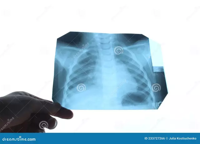
The X-ray picture is polymorphic, as it shows not only manifestations of pneumosclerosis itself, but also concomitant pathology.
Typical is the strengthening and deformation of the pulmonary pattern along the branches of the bronchial tree (with basal pneumosclerosis, the pattern is enhanced in the basal segments of the lungs, with the apical and basal - in the upper parts and the basal zone, respectively), due to deformation of the walls of the bronchi, the pulmonary pattern is mesh and looped. Determined by the reduction of the affected lung in size. To obtain a complete picture, a chest x-ray is performed in two projections - frontal and lateral.
A bacteriological examination of sputum with an antibioticogram, general blood and urine tests are performed.
In order to clarify the diagnosis, computed and / or magnetic resonance imaging can be prescribed.

Source: myshared.ru
Treatment of pneumosclerosis
In the absence of clinical manifestations in active therapy there is no need, the main thing in the treatment of pneumosclerosis in this case is the elimination of etiological factors.
The presence of an acute inflammatory process in the lungs or the development of complications may become an indication for hospitalization of the patient in a pulmonary hospital. At elevated body temperature, patients are shown bed rest.
Drug therapy consists in the use of mucolytic drugs, bronchospasmolytics, immunosuppressive drugs. With circulatory failure, cardiac glycosides are prescribed. With concomitant bronchitis, pneumonia, bronchiectasis, anti-inflammatory and antibacterial drugs are prescribed.
To improve the drainage of the bronchial tree, therapeutic bronchoscopy is performed. At the initial stages of the disease, pneumosclerosis is effectively treated with stem cells.
In patients with pneumosclerosis, the absorption of nutrients is lower, in addition, due to a decrease in oxygen concentration in the blood, the risk of developing gastritis, cholecystitis, and gastric ulcer increases. Therefore, an important link in treatment is diet. The fractional feeding regime is recommended. The diet should be high in calories and at the same time easily digestible. Alcohol, sour, spicy, salty, smoked, fatty foods, and mushrooms are completely excluded. With the development of cor pulmonale, the amount of fluid is limited in order to prevent edema and reduce the load on the heart.
In order to stabilize breathing, physiotherapy exercises (especially breathing exercises and swimming) are indicated, massage of the chest is recommended. Physiotherapy is effective: electrophoresis with drugs, oxygen therapy, diathermy or inductometry on the chest area, ultrasound therapy, ultraviolet irradiation or the use of a Solux lamp.
If large areas of the lung are affected by pneumosclerosis, indications for surgical intervention arise, the atrophied part of the lung must be removed. With severe diffuse changes, lung transplantation may be required.
Possible complications and consequences
Pneumosclerosis can be complicated by arterial hypoxemia, chronic respiratory failure, emphysema, pulmonary heart disease, malignant neoplasms, the addition of a secondary infection (including mycotic, tuberculous origin), patient disability and death.
Forecast
The prognosis depends on the rate of development of heart and respiratory failure. With timely diagnosis and correctly selected treatment, the prognosis is generally favorable.
If complications develop, the prognosis worsens.
Prevention
To prevent the development of pneumosclerosis, it is recommended:
- timely treatment of diseases that can lead to pneumosclerosis;
- quitting bad habits (including avoiding secondhand smoke);
- annual prophylactic fluorography;
- rejection of the irrational use of medicines;
- increased immunity: balanced nutrition, sufficient physical activity, good rest;
- avoiding lung injury.
YouTube video related to the article:

Anna Aksenova Medical journalist About the author
Education: 2004-2007 "First Kiev Medical College" specialty "Laboratory Diagnostics".
The information is generalized and provided for informational purposes only. At the first sign of illness, see your doctor. Self-medication is hazardous to health!


