- Author Rachel Wainwright [email protected].
- Public 2023-12-15 07:39.
- Last modified 2025-11-02 20:14.
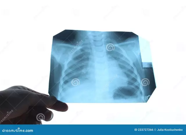
Fluorography of the lungs

Fluorography of the lungs is an examination of the chest organs using X-rays that penetrate the lung tissue and transfer the pattern of the lungs to the film through fluorescent microscopic particles.
A similar study is carried out to persons who have reached the age of 18. Its frequency is no more than 1 time per year. This rule applies only to fluorography of healthy lungs when an additional examination is not required.
It is believed that fluorography of the lungs is not a sufficiently informative examination, but the data obtained with its help can reveal changes in the structure of the lung tissue and become an occasion for further more detailed examination.
The organs of the chest absorb radiation in different ways, so the image looks inhomogeneous. The heart, bronchi and bronchioles look like light spots, if the lungs are healthy, fluorography will display the pulmonary tissue homogeneous and uniform. But if there is inflammation in the lungs, on fluorography, depending on the nature of the changes in the inflamed tissue, either darkening will be visible - the density of the lung tissue is increased, or lightened areas will be noticed - the airiness of the tissue is quite high.
Fluorography of the lungs of a smoker
It has been found that changes in the lungs and airways occur imperceptibly even after the first smoked cigarette. Therefore, smokers - people who are at increased risk of lung disease, are strongly advised to undergo lung fluorography annually.
Not always fluorography of a smoker's lungs will be able to show the development of a pathological process at an early stage - in most cases it starts not from the lungs, but from the bronchial tree, but, nevertheless, such a study allows you to identify tumors and seals in the lung tissue that appeared in the lung cavities fluid, thickening of the walls of the bronchi.
It is difficult to overestimate the importance of passing such an examination by a smoker: pneumonia detected in a timely manner using fluorography makes it possible to prescribe the necessary treatment as soon as possible and avoid serious consequences.
Deciphering a fluorogram after passing lung fluorography

The results of fluorography are usually prepared for several days, after which the received fluorogram is examined by a radiologist, and if a fluorography of healthy lungs was performed, the patient is not sent for further examination. Otherwise, if the radiologist detects changes in the lung tissue, the person can be sent to a X-ray or to an anti-tuberculosis dispensary to clarify the diagnosis.
The image obtained after fluorography of the lungs is accompanied by the conclusion of the radiologist, in which the following wording may appear:
- The roots are widened and compacted The roots of the lungs form lymph nodes and blood vessels, pulmonary vein and artery, main bronchus, bronchial arteries. A lump in this area with a generally satisfactory state of health indicates bronchitis, pneumonia and other inflammatory, possibly chronic processes.
- The roots are heavy. Most often, such a conclusion after the performed lung fluorography indicates bronchitis or another acute / chronic process. Such a change in lung tissue is often found on fluorography of the lungs of a smoker.
- Strengthening the vascular (pulmonary) pattern. The pulmonary pattern is formed by shadows of the veins and arteries of the lungs, and if the blood supply is increased due to inflammation, and this can be bronchitis, and the initial stage of cancer, and pneumonia, it is noticeable on fluorography that the vascular pattern is too prominent. In addition, an increase in the pattern revealed on fluorography of the lungs may indicate problems of the cardiovascular system.
- Fibrous tissue. Discovered connective tissue in the lungs suggests that a person had previously suffered a lung disease. It could have been an injury, infection, or surgery. Despite the fact that such a conclusion indicates the loss of part of the lung tissue, such a result is often given by fluorography of healthy lungs.
- Focal shadows. This is the name of the darkening of the lung area on a fluorogram up to 1 cm in size. If foci are found in the lower and middle parts of the lungs, it may be pneumonia. Severe inflammation is indicated by such a formulation in the conclusion of lung fluorography: "uneven edges", "fusion of shadows", "increased vascular pattern". If the foci are more even and dense, then the inflammatory process is declining. If lesions are found in the upper lungs, this may indicate tuberculosis.
- Calcifications. This is the name for the shadows rounded in shape, reminiscent of bone density. Such phenomena do not pose any danger, but only indicate that the patient had contact with a patient with pneumonia, tuberculosis, infected with parasites, etc., but the body did not allow the infection to develop, but isolated the pathogenic bacteria under deposits of calcium salts.
- Pleuroapical layers, adhesions. Structures of connective tissue, adhesions, found on fluorography of the lungs, in most cases also do not require treatment, but only indicate inflammation in the pleura in the past. Sometimes adhesions cause pain, in which case you should seek medical attention. Pleuroapical layers are called thickening of the apex of the lungs, and they also indicate that a person has suffered inflammation that has affected the pleura (most often tuberculosis).
- The sinus is sealed or free. Pleural sinuses are cavities formed by pleural folds. If the lungs are healthy, fluorography will show that the sinuses are free. But sometimes there is an accumulation of fluid (in this case, treatment is required) or sealed adhesions.
- Aperture changes. Such a conclusion after fluorography of the lungs is given in the event that a person has an anomaly of the diaphragm, which could develop due to poor heredity, obesity, deformation by adhesions, after pleurisy, liver, esophagus, intestine or stomach diseases. In this case, an additional examination is usually prescribed.
- The shadow of the mediastinum is displaced or expanded. The mediastinum is the space between the lungs and the organs in it are the aorta, esophagus, heart, trachea, lymphatic vessels, nodes, thymus gland. The expansion of the shadow of the mediastinum is observed due to an increase in the heart, hypertension, heart failure, myocarditis. Displacement of the mediastinum may indicate uneven accumulation of air or fluid in the pleura, large neoplasms in the lungs. Such a conclusion of lung fluorography indicates that it is necessary to immediately undergo additional examination and treatment.
Found a mistake in the text? Select it and press Ctrl + Enter.


