- Author Rachel Wainwright wainwright@abchealthonline.com.
- Public 2023-12-15 07:39.
- Last modified 2025-11-02 20:14.
Decoding the results of a general blood test
The content of the article:
-
General blood count indicators
- Erythrocytes
- MCV
- Hemoglobin
- Hematocrit
- MCH
- MCHC
- RDW
- Erythrocyte sedimentation rate
- Platelets
- Leukocytes
- How to decipher a blood test
Deciphering the blood test should be done by a qualified specialist, preferably the doctor who wrote out the referral for the study. Despite the presence of reference values of indicators in the form, an independent interpretation of the results often leads to false conclusions.
A general blood test (clinical blood test) is one of the most important laboratory tests in medicine, since the changes occurring in the peripheral blood make it possible to assess the state of the body as a whole.

Blood for general analysis is taken from a finger or from a vein
Usually, blood from a vein is used for analysis, but capillary blood, which is taken from a finger, is also possible. It is recommended to take blood for a general analysis in the morning on an empty stomach; at least eight hours should pass after the last meal. A complete blood count is usually performed using automatic hematology analyzers.
For prophylactic purposes (to assess the state of the body and early detection of possible diseases), children and adults are recommended to take a general blood test once a year.
When deciphering a blood test, one can suspect the presence of certain pathological processes in the body, for example, inflammation. To clarify the diagnosis, additional laboratory and / or instrumental studies are usually required. So, if there is a suspicion of a violation of carbohydrate metabolism, a blood test for sugar (determination of glucose concentration) and other biochemical tests is performed, the oncological process in the body can be determined by studying tumor markers (PSA, CEA, etc.), and in the diagnosis of gastrointestinal pathology, it is important bacteriological research is important (identification of Helicobacter pylori, etc.).
General blood count indicators
Erythrocytes
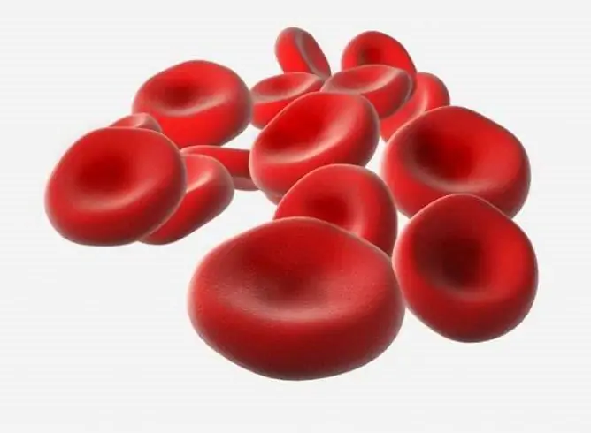
Erythrocytes (red blood cells, RBC, red blood cells) are blood cells that transport oxygen and carbon dioxide. Mature cells do not have a nucleus. The average life span of an erythrocyte is 120 days. In newborns, erythrocytes are slightly larger in size than in adults, which is taken into account when decoding a blood test.

Red blood cells deliver oxygen to tissues
A physiological increase in the number of erythrocytes in the blood is observed in a state of stress, with excessive physical exertion, insufficient nutrition, increased sweating, as well as in children in the first days of life. After prolonged compression of the vein with a tourniquet, falsely increased values may be obtained, which must be taken into account when decoding a blood test.
An increase in the number of erythrocytes in the peripheral blood (erythrocytosis) is recorded with erythremia, absolute or relative secondary erythrocytosis, including cerebellar hemangioblastoma, Itsenko-Cushing's disease, congenital heart defects, chronic lung diseases, burns, ascites, vomiting, diarrhea.
The physiological decrease in the number of red blood cells occurs after eating, between 5:00 pm and 07:00 am, if blood is drawn while lying down.
A decrease in the number of red blood cells (erythrocytopenia) is noted with a lack of iron, vitamins, protein in the body, as well as with hemolysis, leukemia, metastasis of malignant neoplasms.
MCV
In addition to counting the number of red blood cells, a number of morphological characteristics of erythrocytes are also determined, which are calculated by a hematological analyzer or using the appropriate formulas or in a blood smear during the calculation of the leukocyte formula under a microscope.
These erythrocyte indicators include the average erythrocyte volume (MCV). Erythrocytes, whose size is larger than normal, are called macrocytes, less than normal - microcytes. The presence in the blood of red blood cells of different sizes is called anisocytosis. Microcytosis, a condition in which 30-50% of microcytes are detected in the blood, is diagnosed in patients with iron deficiency anemia, thalassemia, microspherocytosis, lead poisoning, and occasionally hyperthyroidism. Macrocytosis, when 50% or more macrocytes are found in the blood, is observed in folic deficiency or B 12 -deficiency anemia, liver disease, smoking and drinking alcohol.
Hemoglobin
Hemoglobin (Hb, HGB) is an iron-containing blood protein whose function is to transport oxygen and carbon dioxide. Hemoglobin is contained in erythrocytes and consists of a protein part - globin and an iron part - heme. Men tend to have higher hemoglobin values than women. In infants, this indicator is physiologically reduced.
An increase in the hemoglobin content occurs with thickening of the blood, pulmonary heart failure, congenital heart defects, diseases that are accompanied by an increase in the number of red blood cells. A physiological increase in the indicator is observed with significant physical exertion, among climbers, residents of high mountain regions, and pilots.
A decrease in hemoglobin in the blood is observed as a result of blood loss during bleeding, increased destruction of red blood cells, a deficiency of iron and / or vitamins in the body, a violation of the formation of blood cells (in the case of hematological diseases). Secondary anemias develop in chronic non-hematological pathologies.
Hematocrit
Hematocrit (Ht, HCT) - an indicator that reflects the proportion of the total blood volume, which is red blood cells.
An increase in hematocrit occurs with erythremia, symptomatic erythrocytosis (respiratory failure, heart defects, kidney tumors), burn disease, dehydration, peritonitis, and shock.
The hematocrit decreases with overhydration, anemia, in the II-III trimesters of pregnancy.
When decoding a blood test, it should be borne in mind that the indicator may slightly decrease when taking blood in the patient's lying position and when taking blood immediately after an intravenous injection, and its false increase occurs with prolonged compression of the vein with a tourniquet.
MCH
The average hemoglobin content in the erythrocyte (MCH) refers to the erythrocyte indices and reflects the synthesis of hemoglobin, as well as its content in the cell (similar to the color indicator, but more accurate). In accordance with it, all anemias are divided into normo-, hypo- and hyperchromic. A false increase in the index is observed with hyperleukocytosis, multiple myeloma. A true increase in MCH occurs with folate deficiency and B 12 deficiency anemias, liver pathologies. A decrease in the index is determined with iron deficiency anemia.
MCHC
The average concentration of hemoglobin in the erythrocyte (MCHC) is the erythrocyte index representing the ratio of the amount of hemoglobin to the volume of the erythrocyte, which reflects the saturation of the cell with hemoglobin. Unlike MCH, this indicator does not depend on the volume of red blood cells.
An increase in the index is observed with spherocytosis, a decrease - with some forms of hemoglobinopathy, iron deficiency anemia.
RDW
The distribution width of red blood cells by volume (RDW) is an erythrocyte index that demonstrates the degree of anisocytosis. An increase in this indicator is observed with a deficiency of vitamins, iron, hemoglobinopathies, severe leukocytosis, hemolytic crisis.
In some cases, it may be necessary to count young red blood cells (reticulocytes), which is usually carried out in a separate analysis.
Erythrocyte sedimentation rate
The blood test takes into account the erythrocyte sedimentation rate (ESR, ESR) - an indicator that reflects the ratio of fractions of blood plasma proteins. A change in the values of this indicator serves as an indirect sign of inflammatory or other pathological processes in the body.
Platelets
Platelets (PLT) are formed elements of blood, small non-nuclear cells of a round or oval shape. Their main function is the formation of a platelet aggregate, which closes the site of damage to the blood vessel, as well as the acceleration of plasma coagulation reactions. On their surface, these elements carry coagulation factors, biologically active substances, circulating immune complexes. Stimulants of platelet aggregation (gluing them together to form a blood clot and stop bleeding) include serotonin, thrombin, collagen, adrenaline.
The number of platelets in the blood varies depending on the time of day and on the season. A physiological increase in platelets occurs after exercise, and a decrease in women during menstruation and pregnancy.
An increase in the number of platelets in the blood (thrombocytosis) is characteristic of systemic inflammatory diseases, anemia, tuberculosis, osteomyelitis, malignant neoplasms, as well as after surgery.
A decrease in the number of platelets (thrombocytopenia) is observed with systemic lupus erythematosus, splenomegaly, congestive heart failure, megaloblastic anemia, disseminated intravascular coagulation syndrome, secondary bone marrow tumors, renal vein thrombosis, against the background of infectious diseases, with massive blood transfusions, and taking certain medications.

Platelets stick together to form a clot that blocks bleeding
The determination of platelets is mandatory during pregnancy, as well as in liver diseases, autoimmune diseases, varicose veins, etc.
A CBC can determine platelet indices such as mean platelet volume (MPV) and platelet volume distribution width (PDW), which are involved in the diagnosis of some diseases.
Leukocytes
Leukocytes (white blood cells, WBC) are blood cells whose main function is to fight infection and tissue damage. Different types of leukocytes (neutrophils, eosinophils, basophils, monocytes and lymphocytes) differ in morphology and functions, their number can change throughout the day.
An increase in the number of leukocytes (leukocytosis) occurs in infectious diseases, inflammatory processes, trauma, and malignant neoplasms.
A decrease in the number of white blood cells (leukocytopenia) is observed in heavy metal poisoning, secondary bone marrow diseases, and some hereditary pathologies.
A detailed blood test involves not only counting the total number of leukocytes, but also drawing up a leukocyte formula, which displays the ratio of various types of white blood cells in the blood and is an important method for diagnosing many pathologies. In particular, characteristic changes in the leukocyte formula make it possible to diagnose leukemia.
How to decipher a blood test
In the result form, as a rule, the name of the indicator (with the corresponding English abbreviation in Latin letters), the obtained and reference values for each indicator is indicated. To correctly read what each value means and to decipher the result of the blood test as a whole, you should contact a qualified specialist - usually it is the doctor who wrote out the referral for the study.
The indicators and their norms necessary for decoding the blood test are shown in the table. In different laboratories, standards and units of measurement may differ.
Table. Decoding a general blood test
| Indicator, units | Reference values |
| erythrocytes (RBC), × 1012 / l |
men - 4-5 women - 3.5-4.7 |
| hemoglobin (HGB), g / l |
men - 130-160 women - 120-140 |
| hematocrit (HCT),% |
men - 42-50 women - 38-47 |
| average erythrocyte volume (MCV), fl | 86-98 |
| average hemoglobin content in erythrocyte, MCH, pg | 27-34 |
| average concentration of hemoglobin in erythrocyte, MCHC, g / dl | 32-36 |
| distribution width of erythrocytes by volume (RDW),% | 11-15 |
| platelets (PLT), × 109 / l | 180-320 |
| leukocytes (WBC), × 109 / l | 4-9 |
| leukocyte formula,% |
segmented neutrophils - 47-72 stab neutrophils - 1-6 eosinophils - 0.5-5 basophils - 0-1 lymphocytes - 19-40 monocytes - 3-11 |
| erythrocyte sedimentation rate (ESR), mm / h |
men - 1-10 women - 2-15 |
YouTube video related to the article:

Anna Aksenova Medical journalist About the author
Education: 2004-2007 "First Kiev Medical College" specialty "Laboratory Diagnostics".
Found a mistake in the text? Select it and press Ctrl + Enter.


