- Author Rachel Wainwright wainwright@abchealthonline.com.
- Public 2023-12-15 07:39.
- Last modified 2025-11-02 20:14.
Trachea
The trachea is an important part of the respiratory tract, connecting the larynx to the bronchi. It is through this organ that air enters the lungs along with the required amount of oxygen.

The trachea looks like a tubular, hollow organ, ranging in length from 8.5 to 15 centimeters, depending on the physiology of the body.
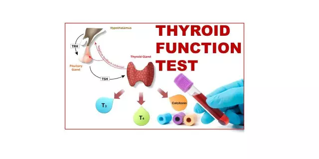
The trachea begins from the cricoid cartilage at the level of the sixth cervical vertebra. One third of the tube is located at the level of the cervical spine, the rest is located in the thoracic region. It ends at the level of the fifth thoracic vertebra, where it divides into two bronchi. In front of the cervical part of the trachea is part of the thyroid gland, and the esophagus is adjacent to the tracheal tube at the back. A neurovascular bundle passes along the sides of the trachea, which includes the fibers of the vagus nerve, the carotid arteries and the internal jugular veins.
The structure of the trachea
If we consider the structure of the trachea in cross-section, it becomes clear that it consists of four layers:
- Mucous membrane. It is a ciliated stratified epithelium lying on the basement membrane. The epithelium contains stem cells and goblet cells, which secrete mucus in small quantities. There are also endocrine cells that produce norepinephrine and serotonin.
- Submucous layer. It is a loose, fibrous connective tissue. This layer contains many small vessels and nerve fibers responsible for blood supply and regulation.
- The cartilaginous part. This layer of the structure of the trachea consists of hyaline incomplete cartilage, occupying two thirds of the entire circumference of the tracheal tube. These cartilages are connected to each other using circular ligaments. In humans, the number of cartilages ranges from 16 to 20. Behind there is a membranous wall in contact with the esophagus, which makes it possible not to interfere with the respiratory process when food passes.
- Adventitia shell. It is presented as a thin connecting shell that covers the outside of the tube.
As you can see, the structure of the trachea is quite simple, but it performs vital functions for the body.
Functions of the trachea
The main function of the trachea is to conduct air to the lungs. However, the number of functions is not limited to this.
The mucous membrane of the organ is covered with ciliated epithelium, moving towards the oral cavity and larynx, and the goblet cells secrete mucus. Thus, when small foreign bodies, such as dust particles, enter the trachea together with the air, they are enveloped in mucus and, with the help of cilia, are pushed into the larynx and pass into the pharynx. Hence, the protective function of the trachea arises.
As you know, warming and purifying the air occurs in the nasal cavity, but the trachea also partially plays this role. In addition, it is necessary to note the resonator function of the trachea, as it pushes air to the vocal cords.
Pathology of the trachea
Conditionally, pathologies can be divided into malformations, injuries, diseases and tracheal cancer.
Malformations include:
- Agenesia is a rare defect in which the trachea ends blindly, without communicating with the bronchi. Those born with this defect are practically unviable.
- Stenosis. It can be obstructive (if there is an obstruction inside the tube) or compression (as a result of pressure on the trachea of abnormal vessels or tumors). In most cases, stenosis is successfully resolved with surgery.
- Fistulas. They are quite rare. They can be incomplete (end blindly) or complete (open onto the skin of the neck and into the trachea).
- Cysts. Have a favorable prognosis for treatment. Surgery is required.
- Diverticula and tracheal dilatation caused by congenital weakness in the muscle tone of its wall.
Tracheal injuries can be open or closed. Closed injuries include ruptures due to trauma to the chest, neck, tracheal intubation. Open injuries include stab wounds, stab wounds, and gunshot wounds.
The most common diseases are:
- Inflammation of the trachea. It can be chronic or acute. As a rule, inflammation of the trachea is combined with bronchitis. Chronic inflammation of the trachea is often a symptom of scleroma, tuberculosis. Inflammation of the trachea can cause fungi Aspergillus, Candida, Actinomyces.
- Acquired stenoses. Distinguish between primary, secondary and compression. Primary stenosis may result from tracheostomy and prolonged tracheal intubation. Physical (radiation damage, burns), mechanical or chemical injuries can also be the cause of stenosis.
- Acquired fistulas. As a rule, they are the result of injuries or the consequence of various pathological processes in the trachea and nearby organs. For example, they can occur as a result of a breakthrough of the peri-tracheal lymph nodes in tuberculosis, opening or suppuration of a congenital mediastinal cyst, with the decay of a tumor of the esophagus or trachea.
- Amyloidosis - multiple submucosal amyloid deposits in the form of tumor-like formations or flat plaques. Amyloidosis leads to a narrowing of the tracheal lumen.
- Tumors. Tumors are primary and secondary. Primary tumors originate from the tracheal wall, and secondary tumors are the result of the invasion of neighboring organs by malignant tumors. There are more than 20 types of benign and malignant tumors. In children, the percentage of benign tumors outweighs (papillomas, fibromas, hemangiomas). In adults, the frequency of benign and malignant tumors is approximately the same. The most common malignant tumors are adenoid cystic cancer of the trachea, squamous cell carcinoma of the trachea, sarcoma, hemangipericytoma. All types of tracheal cancers gradually grow into its wall and go beyond it.
Tracheal intubation
Intubation is the introduction of a special tube into the trachea. This manipulation has a number of technical difficulties, which, nevertheless, are more than compensated for by its advantages in the provision of emergency care to a patient in an extremely serious condition.
Tracheal intubation provides:
- Easy conduction of controlled or assisted breathing;
- Airway patency;
- The best conditions to prevent pulmonary edema;
- The possibility of aspiration from the trachea and bronchi;
In addition, intubation eliminates the likelihood of asphyxia in spasm of the vocal cords, tongue retraction, aspiration of foreign bodies, detritus, blood, vomit, mucus.
The procedure is carried out according to the following indications:
- Terminal state;
- Acute respiratory failure;
- Pulmonary edema;
- Tracheal obturation;
- Severe poisoning, accompanied by respiratory failure.
It is forbidden to do intubation when:
- Any pathological changes in the facial part of the skull;
- Inflammatory diseases of the neck;
- Any damage to the cervical spine.
Found a mistake in the text? Select it and press Ctrl + Enter.


