- Author Rachel Wainwright wainwright@abchealthonline.com.
- Public 2023-12-15 07:39.
- Last modified 2025-11-02 20:14.
Uterus
The uterus (uterus) is an unpaired smooth muscle hollow organ in which the processes of embryo development and fetal bearing take place. The uterus is located in the pelvic cavity, mesoperitoneally, behind the bladder, in front of the rectum. In women of reproductive age, the length of the uterus is approximately 7-8 cm, width - 4 cm. In nulliparous women, the mass of the uterus is 40-50 g, in those who have given birth - about 80 (associated with hypertrophy of the muscular membrane). The uterus is a fairly mobile organ, and depending on the location of adjacent organs, it can take a different position. Normally, the uterus is in the anteflexio position (the longitudinal axis is oriented along the pelvic axis), anteversio (the filled bladder, as well as the rectum, slightly tilt the uterus forward). Most of the surface of the organ, except for the vaginal part of the cervix, is covered with the peritoneum.

The uterus consists of three parts:
- the bottom of the uterus - protrudes slightly above the line of confluence of the fallopian tubes, this is the convex upper part;
- the body of the uterus - the middle part of the conical shape;
- the cervix is a narrowed lower rounded part.
The lower part of the cervix protrudes into the vagina, and is called the vaginal part, the upper part lying above the vagina is called the supravaginal part. On the vaginal part there is an opening of the cervix, which in nulliparous women has a rounded shape, and in those who have given birth it is slit-like.
Layers of the uterine wall
The uterine wall has three layers:
- perimetry (serous layer) - on the greater surface of the anterior, posterior wall and bottom of the uterus is tightly fused with the myometrium, loosely attached to the isthmus;
- myometrium (muscle layer) - consists of three layers of smooth muscles (external longitudinal, middle circular, internal longitudinal) with an admixture of elastic fibers and fibrous connective tissue;
- endometrium (mucous membrane) - formed by cylindrical epithelium, which has superficial (functional) and deep (basal) layers.
Uterus during pregnancy
During pregnancy, the uterus undergoes significant changes. The muscle layer is actively increasing. Muscle fibers increase in length and also become more voluminous. In addition, they increase the content of protein - actomyosin, which is responsible for muscle contractions. To prevent premature contraction of the muscles of the uterus, there is a hormone progesterone. With insufficient production, contractions of the muscular layer of the uterus occur. In this case, we are talking about an increased tone of the uterus. A periodically occurring increase in the tone of the uterus is a variant of the norm, but a constant significant increase in the tone of the uterus can adversely affect the development of the fetus, since when the muscle layer contracts, the blood vessels are compressed, as a result of which the nutrition of the fetus is disturbed. The main danger is inadequate blood supply to the fetal brain. During pregnancy, the uterus increases from the first weeks, reaching its maximum size by the time of delivery.
The muscles of the uterus are always in good shape, not only during pregnancy. They constantly relax and contract. An increase in the tone of the uterus is observed during intercourse, as well as during menstruation, which in the first case contributes to the advancement of sperm, in the second - the rejection of the functional layer of the endometrium.
Erosion of the cervix, treatment
One of the most common diseases of a woman's reproductive system is cervical erosion. Treatment of this pathology is highly effective, but should be carried out on time. The term "erosion of the cervix" means the focus of damage to the mucous membrane of the cervix. Erosion treatment includes the following methods:
- conization;
- laser coagulation;
- chemical coagulation;
- radiosurgical method.
Uterine fibroids, treatment
Another common pathology is uterine fibroids. This is a benign neoplasm that occurs in the myometrium. Myoma is a chaotically intertwining smooth muscle fibers. Fibroid nodes reach quite large sizes, they can weigh several kilograms. Symptoms of this pathology are menorrhagia, pain and a feeling of pressure in the lower abdomen. Symptoms of dysfunction of neighboring organs may also occur: rectum, bladder, arising with large sizes of uterine fibroids. Treatment of this disease can be expectant in nature (this is justified with slow-growing fibroids). In addition to drug therapy, methods such as removal of the uterus, embolization of the uterine arteries, and FUS-ablation of fibroids are used to treat fibroids.
Uterus removal
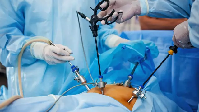
Removal of the uterus, or hysterectomy, is one of the most common surgical interventions in gynecological practice. Removal of the uterus is used for those diseases when the use of alternative methods of treatment is not possible. The indications for this surgery are, in addition to uterine fibroids, endometriosis, uterine prolapse, abnormal uterine bleeding, uterine cancer, cancer of the cervix, ovaries, fallopian tubes.
Depending on the volume of tissue removed, the following types of hysterectomy are distinguished:
- subtotal hysterectomy (amputation of the uterus) - performed with preservation of the cervix;
- total hysterectomy (extirpation) - the uterus is removed with the cervix;
- hysterosalpingo-oophorectomy - the uterus is removed with the appendages;
- radical hysterectomy - the uterus is removed with the appendages, the cervix, the upper part of the vagina, as well as the surrounding tissue, lymph nodes.
Found a mistake in the text? Select it and press Ctrl + Enter.


