- Author Rachel Wainwright wainwright@abchealthonline.com.
- Public 2023-12-15 07:39.
- Last modified 2025-11-02 20:14.
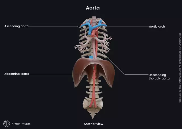
Thoracic aorta
The thoracic aorta is the largest artery in the body that carries blood from the heart.

It is located in the chest, which is why it is called the chest.
The structure of the thoracic aorta
The thoracic aorta is located in the posterior mediastinum and is adjacent to the spinal column. It is divided into two types of branches: parietal and internal.
The internal branches of the thoracic aorta include:
- Esophageal branches, which in the amount of 3-6 are directed to the wall of the esophagus. They branch out ascending branches, anastomosing with the left ventricular artery, and descending, anastomosing with the inferior thyroid artery.
- Bronchial branches, which in the amount of 2 or more branch out with the bronchi. They supply blood to the lung tissue. Their terminal branches approach the bronchial lymph nodes, esophagus, pericardial sac and pleura.
- Pericardial-bursal or pericardial branches, which are responsible for the supply of blood to the posterior surface of the pericardial sac.
- Mediastinal or mediastinal branches, small and numerous, which feed the mediastinal organs, lymph nodes and connective tissue.
The group of parietal branches of the thoracic aorta consists of:
- Posterior intercostal arteries in the amount of 10 pairs. 9 of them pass in the intercostal spaces, from the 3rd to the 11th. The lower arteries lie under the twelfth ribs and are called subcostal. Each of the arteries is divided into a spinal branch and a dorsal branch. Each intercostal artery at the head of the ribs branches into an anterior branch that feeds the rectus and vastus muscles of the abdomen, intercostal muscles, the mammary gland, the skin of the chest, and the posterior branch, which supplies blood to the muscles and skin of the back, and the spinal cord.
- The superior diaphragmatic arteries of the thoracic aorta in the amount of two pieces, which provide blood to the upper surface of the diaphragm.
Arteries of the chest cavity
- Aortic arch;
- Vertebral artery;
- Left and right common carotid arteries;
- The highest intercostal artery;
- Renal artery;
- Aorta;
- Common hepatic artery;
- Left subclavian artery;
- Intercostal arteries;
- Superior mesenteric artery;
- Right subclavian artery;
- Inferior phrenic artery;
- Left gastric artery.
The most common diseases of the thoracic aorta

The most common diseases of the thoracic aorta are aneurysm and atherosclerosis of the thoracic aorta.
Atherosclerosis of the thoracic aorta develops, as a rule, earlier than other forms of atherosclerosis, but for a long time it may not manifest itself in any way. It often develops simultaneously with atherosclerosis of the coronary arteries of the heart or atherosclerosis of the cerebral vessels.
The first symptoms of atherosclerosis, as a rule, appear already at the age of 60-70 years, when the walls of the aorta have already been largely destroyed. Patients complain of recurrent burning pains in the chest (aorthalgia), increased systolic pressure, difficulty in swallowing, dizziness.
Often less specific signs of atherosclerosis of the thoracic aorta are too early aging and the appearance of gray hair, wen on the face, a light strip along the outer edge of the iris, and strong hair growth in the ears.
One of the most dangerous complications of atherosclerosis is aortic aneurysm.
A thoracic aortic aneurysm is a condition in which the weak part of the aorta bulges or expands. The pressure of the blood passing through the aorta leads to its bulging.
Aneurysms pose a serious danger not only to health, but also to the life of the patient, since the aorta can rupture, leading to internal bleeding and death. Up to about 30% of patients with a ruptured aneurysm who are admitted to the hospital survive. This is why a thoracic aortic aneurysm must be treated to avoid rupture.
About half of patients with aneurysms do not have any symptoms of the disease. Most people complain of pain in the lower back and chest, in the neck, back and jaw. Difficulty breathing, coughing, hoarseness occurs.
With a large aneurysm, the aortic valve may be involved, resulting in heart failure.
The most common causes of a thoracic aortic aneurysm are:
- Congenital diseases of the connective tissue (Marfan, Ehlers-Danlos syndrome), cardiovascular system (coarctation of the aorta, heart defects, tortuosity of the aortic isthmus).
- Acquired diseases such as atherosclerosis, or after operations at the sites of aortic cannulation, aortic patches, or anastomotic suture lines of prostheses.
- Inflammatory diseases (infection of the aortic prosthesis, non-infectious and infectious aortritis).
Found a mistake in the text? Select it and press Ctrl + Enter.


