- Author Rachel Wainwright wainwright@abchealthonline.com.
- Public 2023-12-15 07:39.
- Last modified 2025-11-02 20:14.
Neck cyst

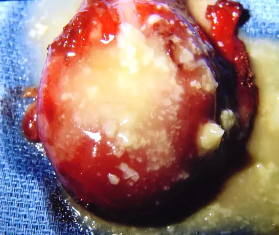
A neck cyst is a tumor-like hollow formation that is located on the lateral or anterior surface of the neck. Contains liquid or slurry. May have complications - malignant degeneration, suppuration or fistula. Neck cysts suppurate in 50% of cases, and fistulas can form after the neck cyst is emptied through the skin.
Types and causes of neck cysts
Neck cysts are median and lateral.
Median neck cysts can sometimes be asymptomatic; they are often found at the age of 4 to 7 years or from 10 to 14 years. Median cysts of the neck are formed as a result of the movement of the rudiment of the thyroid gland along the thyroid-lingual duct from the place of its formation to the front of the neck. This happens at 6-7 weeks of pregnancy.
Lateral cysts of the neck are usually diagnosed at birth. They represent a cavity in the gap between the gill furrows, which should disappear during normal development. The formation of lateral cysts of the neck occurs at 4-6 weeks of pregnancy due to anomalies in the development of the branchial furrows.
Median cysts of the neck
The median cysts of the neck are painless, dense, elastic, not adhered to the skin with clear boundaries, having a diameter of about two centimeters. Located in the midline on the front of the neck and account for approximately 40% of all neck cysts. The median cysts of the neck tend to displace during swallowing, are fused to the hyoid bone and are slightly mobile. Sometimes they can be located in the lingual root. In such cases, the tongue is slightly raised, and swallowing and speech disorders often develop.
Median cysts of the neck are very often suppurate (in 60% of cases). When injected, infections increase in size and become painful. The surrounding tissue becomes red and swollen.
Diagnose median cysts of the neck using ultrasound and puncture of the cyst of the neck with further cytological studies. During the puncture, a viscous, cloudy, yellowish liquid is formed, which contains elements of lymphoids and cells of the squamous stratified epithelium. Diagnosis is based on clinical findings and history. Fistulous passages are examined using probing and fistulography.
The median cysts of the neck are similar to the struma of the tongue, dermoid cysts, lymphadenitis, specific inflammatory processes and adenomas of the abnormally located thyroid gland.
Lateral cysts of the neck

Lateral cysts of the neck are located on the anterior-lateral part of the neck, in its middle or upper third. Localization of the lateral cysts of the neck - next to the internal jugular vein directly on the neurovascular bundle. Lateral cysts of the neck can be either single-chambered or multi-chambered. In cases where they are large, nerves, blood vessels, and also nearby organs can be squeezed.
If there is no compression of the neurovascular bundle or suppuration, the lateral cysts of the neck are not painful. They are an oval or round tumor-like formation, which is easy to notice when the patient turns the head in the opposite direction. Painful sensations are observed on palpation. Above the lateral cysts of the neck, the skin, as a rule, is not changed, and the cysts themselves are mobile, elastic and not adhered to the skin.
With suppuration of the lateral cysts of the neck, they increase in size and become painful. The skin above them turns red, and later a fistula forms.
Lateral cysts of the neck are diagnosed by puncture followed by cytological examinations of the obtained fluid, ultrasound, probing and fistulography with a radiopaque substance. The diagnosis is established on the basis of the clinical picture of the disease and anamnesis.
Uninfected lateral neck cysts are similar to extraorgan neck tumors (lipomas, neuromas) and lymphogranulomatosis. Suppurative - with lymphadenitis and adenophlegmon.
Treatment for neck cysts
Treatment of neck cysts is extremely rapid. Operations on neck cysts are indicated for all lateral neck cysts, as well as for median neck cysts of any size in children and more than one centimeter in diameter in adults. The operation on the neck cyst is performed under intravenous anesthesia, it is removed along with the capsule to prevent relapses. During an operation on a cyst on the neck, the surgeon first makes an incision over the cyst, then he selects and removes the cyst along with the membranes. When removing the median cysts, a part of the hyoid bone is also removed, since the cord from the cyst passes through it. Surgery on lateral cysts of the neck is more difficult because of the nearby nerves and blood vessels.
In elderly patients with concomitant severe diseases, the contents of the neck cyst can be aspirated with further flushing of the cyst cavity with antiseptics. In other cases, this method is not used, since it is not effective enough and has a high risk of relapse.
Due to minimal tissue trauma during operations on neck cysts, the use of modern technology and the imposition of internal cosmetic sutures, patients return to their normal life in the shortest possible time.
The information is generalized and provided for informational purposes only. At the first sign of illness, see your doctor. Self-medication is hazardous to health!


