- Author Rachel Wainwright wainwright@abchealthonline.com.
- Public 2023-12-15 07:39.
- Last modified 2025-11-02 20:14.
Ultrasound of the uterus

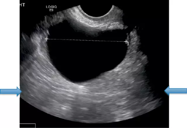
Ultrasound of the uterus and appendages - an ultrasound examination that gives an idea to the gynecologist about the state of the uterus and appendages (fallopian tubes, ovaries), their shape, size, structure.
Based on the results of ultrasound, it is often possible to draw a conclusion about the cause of infertility, diagnose a gynecological disease.
Features of the ultrasound of the uterus and appendages
Ultrasound of the uterus undergoes women who, during a standard examination and on the basis of complaints, showed symptoms of a gynecological disease: pain in the lower abdomen before or during menstruation, prolonged and painful menstruation, intermenstrual bleeding.
In addition, ultrasound of the uterus is performed during pregnancy. The examination is prescribed to confirm the fact of uterine (or ectopic) pregnancy, to exclude cystic drift. Ultrasound of the uterus during early pregnancy helps to find out the state of pregnancy, its duration, and in the presence of pathologies - the essence of the problem and the possibility of prescribing treatment or the need to terminate the pregnancy, A qualitative examination can be carried out only with a full bladder, therefore, a woman is recommended to drink 1.5-2 liters of water a few hours before the ultrasound. An ultrasound scan is performed on the 5-7th day of the cycle.
An ultrasound of the appendages is prescribed to undergo women who have complaints of irregular menstrual bleeding and their absence, pain in the lower abdomen, infertility.
As with ultrasound of the uterus, when examining the tubes and ovaries, if the examination is carried out externally, through the anterior wall of the peritoneum, the woman needs to fill the bladder. If the patient is assigned to undergo a transvaginal examination (the sensor is inserted into the vagina), there is no need to prepare and fill the bladder.
It is advisable to undergo ultrasound of the appendages at the same time as the ultrasound of the uterus. If you need to study the development of ovarian follicles, ultrasound is performed several times during the menstrual cycle.
Results of ultrasound of the uterus
The results of the examination, both abdominal and transvaginal, are received by a woman on her arm shortly after an ultrasound scan, on the same day.

In the conclusion of the doctor, information is provided about the shape of the uterus, its size, the thickness of the walls and endometrium, its correspondence to the day of the cycle, the presence of deviations in development, the structure of the cervix, the size of the ovaries, their structure, the presence of maturing follicles. There should be no data on the fallopian tubes in the ultrasound results of a healthy woman - tubes without pathologies are not visible on the ultrasound.
Norms for the size of the uterus of a non-pregnant woman: length - 70mm, width - 60mm, anteroposterior size - 42mm.
Size standards for ovaries: width - 25mm, length - 30mm, thickness - 15mm.
In addition, the results of an ultrasound of the uterus may contain data on the detected diseases:
- myoma is a tumor of the muscle layer (myometrium) of the uterus of a benign nature. Ultrasound can detect fibroids as small as 1cm in diameter;
- endometriosis. A disease in which the lining of the uterus extends beyond the cavity. With the help of ultrasound, it is impossible to make a final diagnosis, because only small bubbles are visible in the uterine cavity, but the examination makes it possible to prescribe additional tests;
- polyps. They occur when the lining of the uterus expands. On ultrasound, the doctor with this disease notices a thickening or proliferation of the endometrium;
- developmental defects. This is the name for obvious deviations in the size, shape, location of the uterus. Ultrasound of the uterus makes it possible to diagnose a two-horned, underdeveloped, saddle uterus. If a malformation is found, the woman is injected with a contrast for a more detailed study of the organ;
- cervical or endometrial cancer. A neoplasm of a malignant nature found on the cervix or in its mucous membrane;
- polycystic ovary disease. A disease that occurs due to hormonal imbalance. On ultrasound, the doctor notes a thickening of the ovarian capsules, an increase in the ovaries, detects cysts;
- salpingitis. Inflammation of the fallopian tubes, due to which they form adhesions that can cause infertility. The ultrasound shows that the fallopian tubes are thickened.
- tumors of the tubes and ovaries. Ultrasound of the appendages makes it possible to detect tumors, assess their size and structure. For a more accurate diagnosis and treatment, the patient needs to undergo an additional examination.
Found a mistake in the text? Select it and press Ctrl + Enter.


