- Author Rachel Wainwright wainwright@abchealthonline.com.
- Public 2023-12-15 07:39.
- Last modified 2025-11-02 20:14.
Hemoptysis

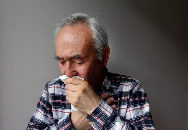
Hemoptysis is the discharge of sputum mixed with blood or a significant volume of blood when coughing from the airways. Blood can evenly stain the sputum brown, red or pink, depending on the disease. In this case, the phlegm can have a foamy or jelly-like appearance. Sometimes an admixture of blood in saliva is mistaken for hemoptysis. Although the sources of blood impurities in saliva can be nosebleeds or bleeding gums.
Causes of hemoptysis
Most often, hemoptysis syndrome is observed with bronchiectasis, tuberculosis, bronchitis, pneumonia, abscess. The causes of hemoptysis can be bronchial adenoma, lung carcinoma, thromboembolism of the pulmonary artery, mitral valve stenosis. Hemoptysis is one of the main symptoms of pulmonary hypertension, pulmonary angiitis, idiopathic progressive pulmonary induration, amyloid dystrophy, and hemorrhagic hemostasiopathy.
The syndrome of hemoptysis and pulmonary hemorrhage can develop when an aortic aneurysm ruptures with its subsequent entry into the bronchus.
With pulmonary tuberculosis, hemoptysis and pulmonary hemorrhage syndrome also often develops. In this case, bleeding is accompanied by pain in the chest associated with inflammation of the pleura, prolonged dry cough of varying intensity and an increase in body temperature.
Regular and prolonged hemoptysis in smokers may indicate the presence of a neoplasm in the lungs.
Diagnosis of hemoptysis
Regular hemoptysis in patients under thirty years of age without signs of another disease indicates a bronchial adenoma. With bronchiectasis, recurrent hemoptysis is accompanied by regular discharge of purulent sputum. Severe pleural pain with hemoptysis indicates a possible myocardial infarction. A physical examination helps to establish the true cause of hemoptysis: a rubbing noise of the serous membrane of the lungs indicates the presence of any pathology associated with damage to the lung membrane (abscess pneumonia, coccidioidosis, angiitis) localized wheezing indicates possible lung carcinoma. The primary examination necessarily includes chest x-ray. But even with normal x-ray results, the likelihood of bronchiectasis or neoplasm as a factor of bleeding remains. A chest x-ray can be used to monitor fluid levels that indicate a build-up of pus or a distal tumor blocking the bronchus. Some patients are prescribed computed tomography of the chest and tracheobronchoscopy. Examination with a rigid endoscope is especially necessary in case of profuse hemoptysis. Examination with a rigid endoscope is especially necessary in case of profuse hemoptysis. Examination with a rigid endoscope is especially necessary in case of profuse hemoptysis.
Help with hemoptysis and pulmonary hemorrhage
Pulmonary hemoptysis is the discharge of large amounts of blood through the airways without or during coughing. Without coughing, blood flows into the mouth from the airways in a stream. The most common causes of pulmonary hemoptysis are lung cancer and tuberculosis.
The blood secreted during pulmonary hemoptysis is scarlet, foaming and does not coagulate. With pulmonary hemoptysis, an emergency hospitalization is indicated.

First aid for hemoptysis is that it is necessary to give the person a half-sitting position, an elevated position, calm him down, prohibit talking and moving. It is strictly forbidden to put cans on the chest, apply mustard plasters, heating pads and hot compresses. An ice pack should be placed on the affected chest area, and the patient should be allowed to swallow small pieces of ice. Reflex spasm when swallowed will decrease the blood supply to the blood vessels of the lungs.
Hemoptysis treatment
The main goal of hemoptysis treatment is to ensure the normal functioning of the lungs and heart and to prevent asphyxia. Treatment of hemoptysis consists in bed rest and taking medications that suppress cough - opiates (dihydroxycodeinone 5 mg four to six times a day, codeine 10-30 mg).
At the very beginning of treatment, the source of bleeding is identified using a rigid bronchoscope, and then the unaffected lung is isolated and ventilated. In case of respiratory failure and massive hemoptysis (the release of about 0.6 liters of blood within two days), resulting from blood entering the respiratory tract, aspiration is required. To isolate the damaged area of the lung, a special tube with an inflatable balloon is inserted to carry out the procedure for incubating the lungs. Taking into account the localization of the source of bleeding and the state of the patient's respiratory function, a classical or surgical method of treating hemoptysis is chosen. Resection of the affected area of the lung should not be carried out in case of inoperable cancer and the expected severe impairment of the external respiration function. In case of significant impairment of lung function, catheterization and embolization of the bronchial artery are performed. In this case, before the procedure, the bleeding area is tamponed with a balloon catheter, lavage is performed with a fibrinogen solution or saline, and vasopressin is injected intravenously.
In case of massive and submassive hemoptysis, the angiography method is used, which includes selective embolization of the bronchial artery. The angiography method allows you to save a significant amount of lung tissue. This method is used for chronic lung disease in patients.
YouTube video related to the article:
The information is generalized and provided for informational purposes only. At the first sign of illness, see your doctor. Self-medication is hazardous to health!


