- Author Rachel Wainwright wainwright@abchealthonline.com.
- Public 2024-01-15 19:51.
- Last modified 2025-11-02 20:14.
Types of ovarian cysts
The content of the article:
-
Classification of ovarian cystic formations
- Follicular cyst
- Corpus luteum cyst
- Paraovarian cyst
- Endometrioid cyst
- Dermoid cyst
- Tecalutein cyst
- Cytadenoma (cystoma)
- Video
Ovarian cysts are benign neoplasms of the pelvis and abdomen. Malignancy (transformation into a malignant tumor) occurs in extremely rare cases. Types of ovarian cysts are associated with the phase of the menstrual cycle in a woman, the presence or absence of pregnancy and the work of the hormonal system as a whole.
These formations are often cystic in nature, less often denser structures appear.

Ovarian cysts differ in origin, structure, size and prognosis
Classification of ovarian cystic formations
There are several main classifications of ovarian cysts. Depending on the time of occurrence:
- Functional cysts. This is a temporary variant of cysts (exist over several menstrual cycles). The reason for the occurrence is in violation of the normal course of menstruation and, in particular, the ovulation process. They are not dangerous and disappear spontaneously, no treatment is required.
- Anatomical cysts. Their occurrence is often associated with an abnormal structure of the ovaries (congenital tissue defects). In rare cases, large functional cysts precede their appearance. In this case, the destruction of the normal structure of the ovaries occurs. Requires medical or surgical treatment.
Depending on the size, there are:
| View | The size | Features: |
| small | up to 5 cm | Only require supervision |
| average | from 5 to 10 cm | Require drug treatment and ultrasound monitoring |
| big | more than 10 cm | Require surgical treatment |
As a rule, all types of ovarian cysts are asymptomatic. In the event of complications (torsion of the leg, rupture), symptoms of an acute abdomen arise, which require emergency hospitalization and treatment in a hospital, and in 90% of cases, surgery is required.
Follicular cyst
Refers to the type of functional neoplasms and is often determined by chance by ultrasound. Only instrumental diagnostics can make a diagnosis, since it rarely manifests itself clinically.
Features of the disease:
- It occurs at a young age.
- It is associated with a violation of ovulation (the follicle does not burst and the egg does not leave it). The follicle cavity begins to fill with fluid, resulting in an enlargement.
- Has a direct connection with the disruption of the hormonal system.
- Rarely reaches large sizes. More often diagnosed in the range of up to 4-6 cm (it can reach up to 12 cm).
The clinical picture occurs when the size of the cysts is more than 8 cm, since in this case there is a high risk of torsion and rupture with the development of a clinic of peritonitis (pain, intoxication, cardiovascular disorders).
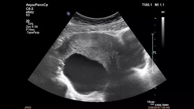
The structure of the neoplasm:
- single chamber;
- single;
- rounded with clear contours;
- thin-walled;
- the contents are homogeneous.
There are no histological features.
Corpus luteum cyst
The corpus luteum is formed in the ovaries under the action of the pituitary luteinizing hormone. The formation of a cystic cavity is based on impaired blood circulation and lymph circulation, which leads to the accumulation of serous fluid. The corpus luteum cyst also belongs to functional formations.
Features:
- It is formed by the gland of temporary secretion (the corpus luteum forms and involutions monthly).
- It occurs at a young age (not typical for menopause, since ovulation does not occur).
- The name is determined by the lipochromic pigment in the gland cavity.
- It is a hormone-producing formation - it releases progesterone.
- It occurs during ovulation and ensures the implantation of the ovum into the endometrium.
- During pregnancy, the regression process occurs at 1-12 weeks.
In rare cases, the cyst cavity can be filled with hemorrhagic contents. The diameter usually does not exceed 6-8 cm. It is found by chance on ultrasound. The clinical picture of the disease occurs extremely rarely, only in the case of complications.
Features of the structure:
- has a cellular structure;
- wall thickness reaches 2-4 mm;
- good blood supply to surrounding tissues;
- has a round or oval shape;
- the contents are homogeneous (echo signs of microbleeds may be added to the cyst cavity).
The histologically homogeneous structure is represented by the cells of the corpus luteum, which are located in several layers.
The cyst goes through several stages of development:
- Proliferation.
- Vascularization.
- Flourishing.
- Reverse development.
Depending on the phase, according to ultrasound data, cysts differ somewhat in size, density and content.
Paraovarian cyst
It arises from the retention formation - epiophoron. This is the embryonic remnant of the primary kidney that lies between the ligaments of the uterus, the ovary and the fallopian tube. It does not leave directly from the tissues of the ovary, but is located in nearby tissues.
Features:
- Large sizes (up to 15-20 cm).
- They arise in adulthood.
- Clinically expressed by pain and pulling sensations in the lower abdomen.
- Affects nearby organs (bladder - causes frequent urination, rectum - false urge to defecate).
- There is a high risk of torsion and rupture, which is why there are urgent indications for hospitalization.
- Immobile.
- Does not regress on its own.
The clinic occurs in 40-50% of women with this pathology. Detection by ultrasound or gynecological examination.
Cyst structure:
- oval or round shape;
- the structure is homogeneous;
- thin-walled (up to 3 mm);
- localized in the immediate vicinity of the ovary, sometimes soldered to its wall.
The histological structure is heterogeneous and is represented by different types of cells.
Endometrioid cyst
This type of cystic neoplasm arises from the endometrium in the ovaries. The endometrium is the inner muscle layer of the cells that line the uterus, but for a number of reasons, some cells can migrate down the fallopian tubes all the way to the ovaries.
During menstruation, some of the cells that should have separated and come out accumulate and a capsule forms around them.
Features:
- Directly related to the menstrual cycle.
- Often there is a clinical picture of the disease in the form of pulling pains, discharge, less often there is an asymptomatic course.
- They have a tendency to malignancy (endometrioid cancer).
- Often accompanied by endometriosis of the uterus and other organs.
- Complications in the form of adhesive disease are frequent.
There are several stages of ovarian endometriosis:
| Stages | Cystic lesion size | Localization and characteristics |
| Stage I | Not formed | There are only point foci of endometriosis on the ovaries (superficial, small diameter). The peritoneum and rectal cavity may be involved. |
| Stage II | Size 5-6 cm | Located on one ovary. Endometriosis foci on the peritoneum. The beginning of the development of the adhesive process in the uterine appendages. |
| III stage | Size 6-7 cm | The defeat involves both ovaries. Foci of endometriosis in the serous layer of the uterus, on the fallopian tubes. Extensive damage to the peritoneum and a pronounced adhesive process. |
| Stage IV | More than 7 cm | Severe damage to both ovaries (loss of organ functionality) and total damage to surrounding organs and tissues. |
Features of the structure:
- dense bluish capsule;
- the content is chocolate or black and gray;
- on the surface there are multiple foci of endometriosis (erosive surfaces);
- the content is inhomogeneous;
- contours are often uneven;
- can be both single-chamber (less often), and have multiple constrictions (divides the cavity into multiple chambers).
The histological structure is heterogeneous, contains cells of the uterus, ovaries, blood clots.
Dermoid cyst
Dermoid cysts are benign formations and are relatively less common than others. They arise from epithelial tissue, so they can contain hair, follicles, sebaceous and sweat glands.
Features:
- Large sizes (up to 15 cm).
- Complications are rare.
- In 80%, they are not clinically manifested.
- Slow growth.
- They meet at a mature and young age.
- Only one-sided defeat.
It is found during an ultrasound scan and during a gynecological examination. If the diagnosis is difficult or for differential diagnosis, it is necessary to perform computed or magnetic resonance imaging.
Features of the structure:
- heterogeneous structure;
- thick dense capsule;
- the contours are uneven;
- are both single-chamber and multi-chamber.
Histologically heterogeneous structure, because it contains cells of many different tissues.
Tecalutein cyst
Cystic neoplasms arise from atresized follicles under the influence of excessive function of gonadotropic hormones of the pituitary gland, sometimes due to trophoblastic disease (cystic drift).
Features of the disease:
- There are clinical signs.
- There are concomitant pathologies from the reproductive system (menstrual irregularities, infertility).
- Bilateral defeat.
- Large size (up to 30 cm).
- High risk of complications.
- Abdominal effusion is often found, which can mimic an acute abdomen clinic.
- Detection by ultrasound, clinical picture and gynecological examination. Sometimes they occur as a result of ovarian hyperstimulation during IVF treatment for infertility.
Features of the structure:
- multi-chamber;
- round or oval shape;
- there are constrictions on the surface, which divides the cyst into lobules;
- the walls are smooth and thin;
- the contents are homogeneous (more often serous, less often there is an admixture of blood).
The histological picture is represented by atresized follicles and a layer of luteinized cells.
Cytadenoma (cystoma)
It is not a classically cystic formation, since it occurs due to cell division, and not due to the accumulation of fluid. It is considered in the context of cysts, since it has a cavity with contents.
Features of the tumor:
- It occurs in women after menopause (the period of extinction of sexual function).
- Clinically manifested by pulling pains, impaired urination and defecation.
- Considered in the context of a precancerous condition (frequent malignancy).
- Large sizes (up to 15 cm).
There are two types of formations:
- Serous cystadenomas - formed by the surface layer of the ovarian epithelium, a clear liquid is contained inside the cavity.
- Mucinous cystadenomas - formed by the same, but the contents are cloudy and viscous, slimy.
Tumor structure:
- there are both single-chamber and multi-chamber;
- the content is inhomogeneous and depends on the type of education.
The histological picture is presented by abnormal (tumor) cells.
Video
We offer for viewing a video on the topic of the article.

Anna Kozlova Medical journalist About the author
Education: Rostov State Medical University, specialty "General Medicine".
Found a mistake in the text? Select it and press Ctrl + Enter.


