- Author Rachel Wainwright wainwright@abchealthonline.com.
- Public 2024-01-15 19:51.
- Last modified 2025-11-02 20:14.
Mole on the head
The content of the article:
- What does it look like
-
Red mole on the head: is it dangerous
- Hazard criteria
- What should a patient do if criteria are met
- Who is at risk
- How is the appointment with the doctor
- Mole on the head in the hair: what is the reason for the appearance
-
Treatment
- What to do at home
- Surgery
- Video
Moles (nevi) and birthmarks are benign growths that can appear anywhere on the body and in any quantity. In most cases, they are not dangerous to human health and life. A mole on the head can be dangerous when it is often injured or changes its color, shape, size. In this case, you need to see a doctor, undergo an examination and, possibly, remove the formation.

Most moles on the head are not dangerous.
What does it look like
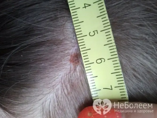
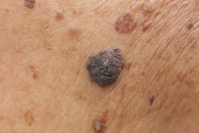
The appearance of education in the photo may vary. All of these options are normal:
- The color is most often brown, but can be black, red and even purple. A white border may appear around.
- Raised (convex) or not raised (flat) above skin level.
- A mole can be located on any part of the head: scalp (back of the head, crown), face.
- The size ranges from spot to 5 cm and even more. Birthmarks are more common.
- There are no additional symptoms. The patient should not be disturbed by pain, numbness, burning sensation. The appearance of these symptoms most likely means that we are talking about another pathology.
Red mole on the head: is it dangerous
In most cases, moles on the head are not dangerous and do not require diagnosis or treatment. However, there is concern associated with the likelihood of malignancy (malignancy) of such formations. Under the influence of various factors (damage, ultraviolet radiation), a mole can degenerate into melanoma. That is why every person should be able to distinguish between a dangerous mole according to special criteria.
Hazard criteria
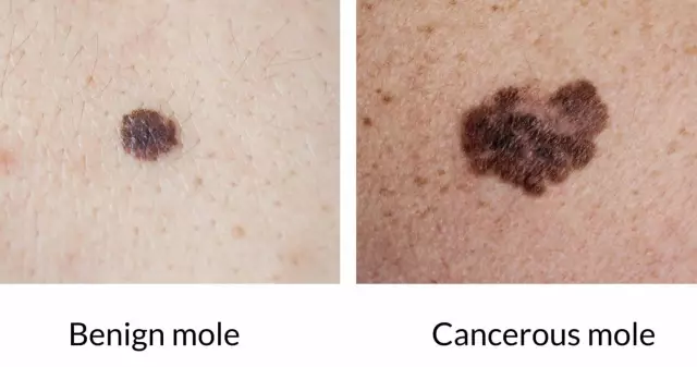
Moles on the scalp require special attention, as they are often injured when combing or other manipulations. There are 5 criteria that help to assess the danger of a nevus: asymmetry, edge, color, size, dynamics (abbreviated as ACORD).
| Criterion | Explanation |
| Asymmetry | If you draw a conditional line in the center of the formation, both halves should be the same, even, symmetrical. Asymmetrical halves of a nevus are one of the signs of danger. |
| Edge | Normally, the edge of the nevus is smooth and clear. Blurred edges, as well as jags, deformation are a sign of danger. |
| Coloration | Education can have a different color: brown, black, red (all these options are the norm). It is not the color itself that is important, but its uniformity. The presence of any inclusions, darkening, blanching should alert the patient. |
| The size | The larger the nevus, the higher the risk of malignancy. The diameter of the formation is more than 6 mm - a warning sign. |
| Dynamics | It is very important to monitor the nevus over time. The appearance of roughness, ulceration, as well as the rapid growth of education should be alerted. |
If the formation is localized on the head, it is not always possible to conduct a self-examination. It is better to ask someone close to you to examine hard-to-reach places (the back of the head, crown).
What should a patient do if criteria are met
If at least one of the above criteria is identified, you must definitely contact a dermatologist. This does not always indicate a malignant nevus, but it is better to play it safe.
Who is at risk
There is a group of people who are especially at risk of degenerating nevus into melanoma. The risk group includes:
- men over 50;
- persons with a burdened heredity (melanoma in relatives);
- persons who have more than 100 moles;
- persons subject to sunburn;
- faces with fair skin, red hair, freckles.
Anyone at risk of melanoma should be seen regularly by a dermatologist. The frequency of preventive examinations is once a year (even without dangerous signs).
How is the appointment with the doctor
The doctor will carefully examine the nevus, determine the presence of signs of malignancy, and take a history of life and disease. An additional examination is prescribed, usually the consultation is limited to dermatoscopy. Dermatoscopy is a visual assessment of skin growths. For the study, a dermatoscope is used, with which you can get an image magnified 10 times.
Dermatoscopy is a preliminary diagnostic method. It will not be possible to make a final diagnosis (benign or malignant formation, what kind), for this a histological examination is necessary.
Mole on the head in the hair: what is the reason for the appearance
A mole is a collection of melanocytes that are located between the lower (dermis) and upper layers of the skin (epidermis). Melanocytes are skin cells that produce the pigment melanin. It is this pigment that ensures the color change of moles. Red formations represent the proliferation of blood vessels.
Almost every person has birthmarks. They can be formed under the influence of various factors - genetic, hormonal, external environment.
| Cause | Value |
| Heredity | One of the most common reasons for the formation of nevi (both congenital and acquired) is genetic abnormalities. Environmental factors are superimposed on the hereditary predisposition. |
| Hormones | Nevi grow under the influence of various hormones. For example, the number of moles increases during puberty, in women during pregnancy. |
| Ultraviolet radiation | Sunlight triggers the production of melanin, which leads to the formation of moles. |
| Other factors | Injuries, viruses, radiation can provoke the movement of melanocytes to the epidermis. |
Treatment
Not all nevi can be removed. In most cases, it is enough to follow simple guidelines for preventing complications. If a mole is often injured or there are signs of malignant degeneration, such a formation must be removed.
What to do at home
All measures taken at home are aimed at preventing complications. No home treatment allows you to qualitatively remove the formation.
Care Tips:
- try to gently wash and comb your hair so as not to damage the nevus;
- do not use scrubs and peels on the scalp;
- if the mole is located on the open part of the head, cover it with a sun bandage.
One of the most important things you can do at home is checkups. It is necessary to regularly examine the nevus and evaluate it according to the ACORD algorithm.

Only a doctor should remove moles in appropriate conditions, self-medication is unacceptable
Surgery
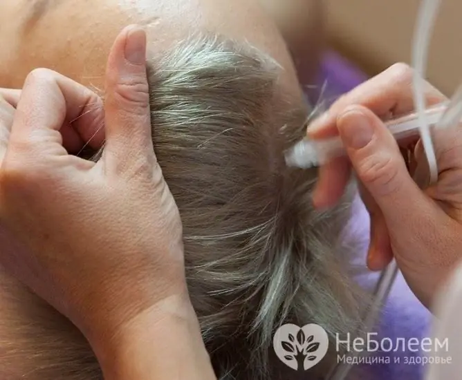
The patient should make the final decision on the need to remove the nevus together with the doctor after a full examination. Surgical treatment is indicated for all patients with suspected melanoma. Various methods can be used to remove the formation - laser therapy, electrocoagulation, cryodestruction, surgical excision.
| Treatment method | Description, advantages and disadvantages |
| Laser therapy | Suitable for removing nevi that are often traumatized (especially on open head areas). This is a modern method of treatment, a laser is used to remove the formation. The procedure is carried out under local anesthesia; no scars remain at the site of removal. The main disadvantages are the high cost of treatment and the inability to remove malignant tumors. |
| Electrocoagulation | The method is based on exposure to electric current. Cauterization of the nevus occurs, after a while the necrotic tissues are rejected on their own. The procedure is performed under local anesthesia. |
| Cryodestruction | Exposure to liquid nitrogen is often used to remove small formations. Freezing and subsequent destruction of the mole occurs. The procedure is painless, does not require anesthesia and local anesthesia, but is not suitable for removing large nevi. Sometimes it takes multiple sessions to completely remove. |
| Surgical excision | Surgical excision is the main treatment for melanoma and large benign neoplasms. A scalpel is used for removal, the operation is performed under local or general anesthesia. After excision of a nevus or melanoma, the wound is sutured, and the formation is sent for histological examination. |
Video
We offer for viewing a video on the topic of the article.

Anna Kozlova Medical journalist About the author
Education: Rostov State Medical University, specialty "General Medicine".
Found a mistake in the text? Select it and press Ctrl + Enter.


