- Author Rachel Wainwright [email protected].
- Public 2023-12-15 07:39.
- Last modified 2025-11-02 20:14.
Features of CT and MRI methods
Every adult has at least once faced such a medical manipulation as hardware diagnostics. This can be an ultrasound or X-ray, an annual fluorographic examination, or an EKG. However, not everyone is aware of the tomographic methods of remote scanning - CT and MRI. This is due to the relative high cost of these procedures, the availability of which is increasing every year.

How do tomographic scanning methods work?
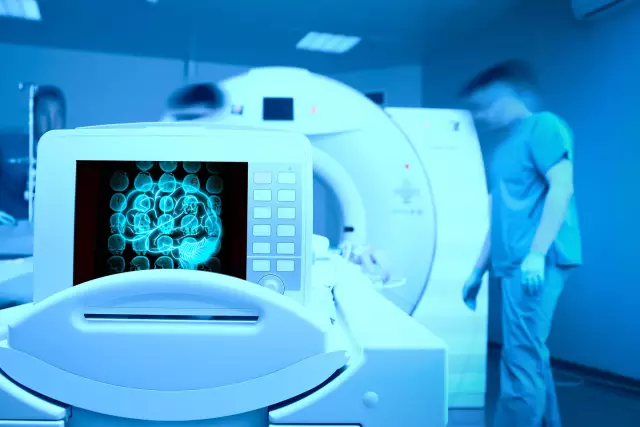
Screenings carried out on hardware devices - tomographs are carried out according to a single plan: the patient is placed inside the machine, remains motionless throughout the entire process, the scanner takes a layer-by-layer survey of the area under study and transfers the images to a computer monitor. The images are obtained in black and white colors with more than 500 shades of gray, which are differentiated by the equipment and recognized as pathological foci.
Despite the external similarity, these types of surveys differ significantly. CT and MRI have different operating principles. Thus, computer screening uses ionizing radiation identical to an X-ray machine. Biological structures are "translucent", dense, cavity or liquid objects are seen on scans more clearly than soft tissue formations. Therefore, computer screening is more often used to examine skeletal structures, dynamic hollow organs (heart, lungs), vasculature, and abnormal neoplasms.
Research on an MRI machine does not involve the use of X-rays, as it is carried out on the basis of capturing signals from the molecular structures of biological cells. When a patient is placed inside a tomograph, his body is exposed to a high-frequency magnetic field. Atoms of cells enter into dissonance with the external field of the apparatus, emit certain signals that are recorded by the equipment. This is the main difference between CT and MRI. Since the structure of soft tissues is more "free" in comparison with dense formations and systems, the internal resonance from them is higher. The MRI images show more clearly soft tissue organs, brain structures, nerve canals, muscle and tendon fibers, cartilage, etc.

Where is the diagnosis carried out?
Any research medical centers equipped with the necessary equipment accept * CT and MRI *. So far, not all district polyclinics are equipped with high-precision scanners, but the study is available for passage in any area of the capital. To search for the nearest medical organization, you can use the unified city recording service https://mrt-v-msk.ru/kt/. Since the clinics on the site are listed, it makes it easy to find and compare the required institution by address, rating and prices for tomography. Follow the link, choose the best offer and sign up for the procedure immediately through the portal. This will make it possible to take advantage of a discount of 1000 rubles for any type of diagnosis.
Found a mistake in the text? Select it and press Ctrl + Enter.


