- Author Rachel Wainwright [email protected].
- Public 2023-12-15 07:39.
- Last modified 2025-11-02 20:14.
Metacarpal bone
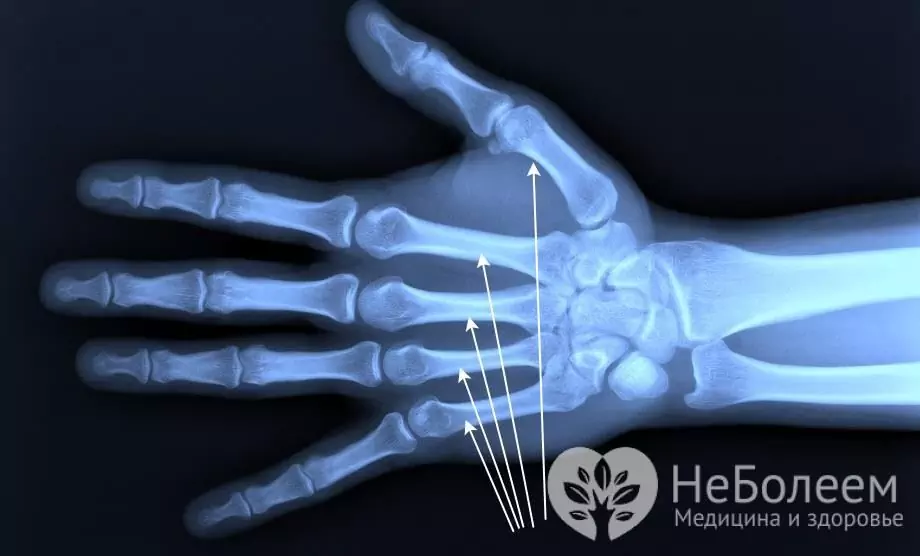
The metacarpal bone is a short tubular bone located on the hand and extending from the wrist in the form of a ray. A person has five metacarpal bones on each hand. Each bone consists of a base, body, and head. These bones are connected by joints to the bones of the wrist and the base of the first phalanx of the fingers.

The structure of the metacarpal bone
The metacarpal bones are measured from the thumb and are curved towards the hand. Each such bone has a body and pineal gland. The body of the metacarpal bone has three surfaces - posterior, medial and lateral. The medial and lateral surfaces are separated by a ridge, where there is an opening leading into the nutrient canal.
The body of the metacarpal bone is concave dorsally, and the lateral surfaces of the base are the articular areas that connect the adjacent bones. The articular surfaces are saddle-shaped.
The base of the third metacarpal bone has a styloid process. At the bottom of the distal part is the spherical head of the metacarpal bone. The lateral surfaces of the metacarpal head are rough.
Each metacarpal head and body can be felt through the skin on the surface of the hand. Between the metacarpal bones there are interosseous spaces called the metacarpals.
Metacarpal injuries
The most common injuries are fractures of the metacarpal bone, base, shaft, and phalanges. The most common fracture occurs in the first and fifth metacarpal bones. Injury can be caused by direct impact with a blunt object.
In rare cases, fractures of the second, third and fourth metacarpal bones occur. Typically, this fracture occurs due to an injury to the hand or a blow with a fist on a blunt object.
Fractures of the metacarpal bone at the base are of several types: intra-articular, extra-articular and transverse. Symptoms are pain in the area of the fracture, edema, inability to bend the finger, and when probing the site of the fracture, the pain syndrome increases. Bennett's fracture is an injury in which there is a triangular-shaped splinter, as well as a dislocation towards the radius. Complicated fracture with dislocation is called Roland's fracture. An accurate diagnosis is made with X-ray examination.
Treatment of a base fracture begins with local anesthesia and a plaster cast over the fracture site. In case of serious damage and the presence of fragments, surgical intervention is performed. A plaster cast is applied for five weeks, and after it is removed, the patient is assigned physical therapy and physiotherapy.
Rarely is a metacarpal shaft fracture that occurs with or without displacement. Symptoms are pain in the area of injury, severe stress, and displacement of the first toe.
Treatment begins with an X-ray and a plaster cast from the forearm to the base of the fingers. In some cases, surgical treatment and finger fixation with needles are required.
Fracture of the phalanges of the fingers occurs with a strong direct or indirect blow to the finger. Such a fracture has several types: transverse, helical, comminuted, intra-articular and extra-articular. Symptoms include pain, swelling of the hand, swelling of the finger, and soreness when extending the hand. At the first examination, a deformation of the finger is observed.
Treatment begins with matching the broken bone fragments and returning the phalanx to its normal position. A plaster splint or splint is applied to the finger for 30 days. In case of serious injuries, the finger is fixed with knitting needles and a bone pin, and then a plaster cast is applied.
Found a mistake in the text? Select it and press Ctrl + Enter.


