- Author Rachel Wainwright [email protected].
- Public 2023-12-15 07:39.
- Last modified 2025-11-02 20:14.
Elbow bone

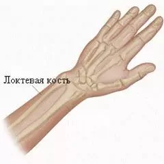
The ulna consists of a body and two epiphyses - distal and proximal. The body of the bone has a triangular shape, it has three edges: palmar (anterior), external (interosseous) and dorsal (posterior), and three surfaces.
The structure of the ulna
The anterior edge of the bone has a rounded shape, the posterior edge is directed backward, and the interosseous edge has a pointed shape and faces the radius. On the ulna there is a feeding opening leading to the proximal feeding tubule. On top of the front surface of the bone, on the border between the upper end and the body, there is a tuberosity. The posterior surface of the bone is turned back, and the medial surface is turned towards the inner side of the forearm.
The proximal pineal gland has a slightly thickened shape and at the top passes into the olecranon process. In front, this process is occupied by a block-shaped notch, which is bounded on the lower side by the coronoid process. On the outer part of the coronoid process, the ulnar notch is located - the junction of the articular circumference of the radial head with the ulna. From the back of the radial notch, the crest of the instep support begins, which goes down and reaches the upper edges of the bone body.
The distal pineal gland has a slightly rounded shape. On it you can clearly see the head of the ulna. Its surface is concave and smooth, facing the wrist. Along its periphery is the articular surface - the articular circumference of the bone, which connects to the radius. The medial-posterior surface of the head passes into the styloid process, which is easily felt through the skin.
Ulna fractures

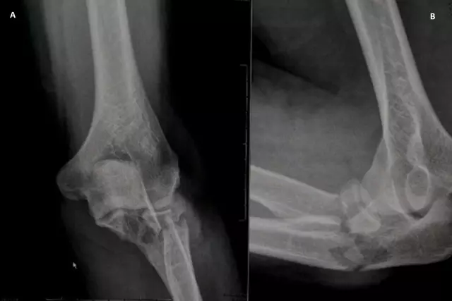
Often, a fracture of the ulna at the border of its middle and upper third or in the upper third is accompanied by a dislocation of the radial head, which can move forward and upward, or upward and outward.
Thus, displacement of the radial head after a fracture of the ulna often leads to damage to the branch of the radial nerve. Such damage often occurs as a result of a strong direct blow, for example, with a stick on the forearm, extended forward and upward. As a result of such a blow, a bone fracture occurs with a significant displacement of the fragments at an angle, from the back side open to the ulnar side, and as a result, a dislocation of the radial head occurs. This injury, typical of a defending person trying to ward off a blow to the head, is called a Montage injury or parry fracture.
As a result of the fracture, the posterior contour of the forearm is severely bent, and the elbow joint expands as a result of the dislocation of the head. During palpation, the patient feels a sharp pain, and the head of the radius protrudes.
Any movements in the elbow joint (passive, active, supination, pronation) are accompanied by severe pain and are therefore limited. With passive flexion in the elbow joint, springy movements are observed.
Found a mistake in the text? Select it and press Ctrl + Enter.


