- Author Rachel Wainwright [email protected].
- Public 2023-12-15 07:39.
- Last modified 2025-11-02 20:14.
Keratoacanthoma

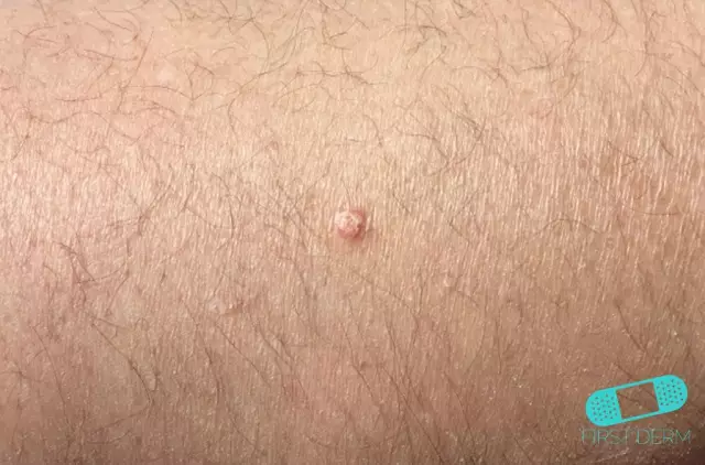
Keratoacanthoma is a benign tumor that often recurs after cryodestruction and is located on the face, exposed parts of the body, forearms and limbs, mainly in old age. Rarely, swelling occurs on the palate, cheeks, lips, nose, or under the nails.
In 6% of cases, keratoacanthoma, if untreated, flows into squamous cell carcinoma. Keratoacanthoma of the skin has the appearance of a domed exophytic node from a grayish-red to blue tint. The central part of the tumor is filled with horny masses, and at the edges it rises above the level of the skin. Keratoacanthoma grows rapidly, reaching large sizes within a few days or weeks.
Classification of keratoacanthoma
Keratoacanthomas are divided into the following types:
- Giant (up to 10-20 mm in size);
- Solitary (single);
- Keratoacanthomas with peripheral growth;
- Subunguals, which grow from the nail bed to the nail plate at a rapid rate and gradually become crusty nodes;
- Multiple in combination with immunosuppression and Togge's syndrome;
- Mushroom, having the appearance of flat or convex tumors with a smooth surface, which is covered with orthokeratosis masses;
- Multimodular, which is characterized by the presence on the tumor of several confluent or isolated craters with the formation of large irregular ulcers;
- Tubero-serpiginous, having the appearance of a hemispherical focus of irregular shape, with thinned skin and a small amount of horny masses with central ulceration.
Causes of keratoacanthoma
The causes of keratoacanthoma are:
- Smoking;
- Ultraviolet irradiation;
- Exposure to tar, carcinogens and mineral oils;
- P53 gene mutations;
- Decreased immunity;
- Radiation;
- Mechanical injury;
- Genetic predisposition;
- Viral infections (papilloma virus and others).
Development of keratoacanthoma of the skin
In its development, the tumor goes through several stages:
- At the first stage, keratoacanthoma is in the stage of active growth, which begins with the appearance of a small and gradually growing tubercle;
- At the second stage, the tumor stops growing and stabilizes;
- At the third stage, a phase of sudden regression begins, when the tumor disappears, and a scar forms in its place.

The third stage usually occurs 6-9 months after the onset, and sometimes never occurs without surgery. That is, either the tumor will disappear, leaving behind a scar, or it will turn into squamous cell carcinoma.
Diagnostics of the skin keratoacanthoma
Diagnosis of a tumor is based on clinical manifestations, by means of excisional biopsy and biopsy of the cushion area. Based on the studies carried out, the stage of keratoacanthoma and its further treatment are determined.
Keratoacanthoma treatment
In many cases, keratoacanthoma of the skin goes away on its own, without requiring any medical intervention, but the consequences in the form of scars do not disappear on their own. This fact and the ability to develop into malignant tumors are the main reasons for the surgical treatment of tumors.
Surgical treatment of skin keratoacanthoma can occur with the help of:
- Laser therapy, which removes growths on any part of the skin;
- Cryosurgery, which is effective in the early stages of the tumor and for which liquid nitrogen is used;
- Electrosurgery, which is performed by electric current;
- Cutting off the tumor with a surgical scalpel.
YouTube video related to the article:
The information is generalized and provided for informational purposes only. At the first sign of illness, see your doctor. Self-medication is hazardous to health!


