- Author Rachel Wainwright [email protected].
- Public 2023-12-15 07:39.
- Last modified 2025-11-02 20:14.
Granuloma

Granuloma is a limited accumulation of cells of young connective tissue, resembling a small nodule in appearance. Such formations appear when the body is damaged by various infectious agents (tuberculosis, syphilis, rabies, etc.) or collagen diseases (for example, rheumatism). In addition, they develop at the site of a foreign body entering the skin and mucous membranes.
Origin of granulomas
The etiology of granulomas is diverse. One of the classifications of these formations is based precisely on their origin:
- Non-infectious;
- Infectious;
- Unidentified genesis.
Non-infectious granulomas appear as a result of drug exposure (granulomatous hepatitis) or occupational dust diseases (talcosis, silicosis, asbestosis, etc.). They also develop around various foreign bodies.
The treatment of granulomas of an infectious nature is closely related to the origin (with typhoid and typhus, tularemia, rabies, syphilis, viral encephalitis, tuberculosis, etc.), since you can get rid of small foci of inflammation only by destroying the pathogen that caused them.
Granulomas of unknown origin include neoplasms in Horton and Crohn's diseases, sarcoidosis, Wegener's granulomatosis.
Types of granulomas
In the classification by morphological characteristics, there are three main types of granulomas:
- Epithelioid cell, or epithelioidocytoma;
- Macrophage, or phagocytomas;
- Giant cell.
According to the level of metabolism, formations with a high and low level of metabolism are distinguished. The first appear under the influence of toxic agents (leprosy, mycobacterium tuberculosis, etc.) and are epithelioid-cell nodules. The latter arise under the influence of inert bodies and consist of giant cells of foreign substances.
According to another classification, granulomas are divided into two groups:
- Specific, the morphology of which is characteristic of a specific infectious disease. When researching, the pathogen can be isolated from the overgrown cells. This group includes annular, leprosy, tuberculous, scleroma, syphilitic granulomas;
- Non-specific, without any characteristic features. They arise both in infectious diseases (typhus and typhoid fever, leishmaniasis) and in non-infectious diseases (silicosis, asbestosis).
The features of the disease can be considered using the example of two interesting varieties of granulomas: eosinophilic and pyogenic.
Pyogenic granulomas (botryomycomas)
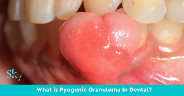
They are slightly raised formations of scarlet, brown or blue-black color. The growth of pyogenic granulomas is caused by edema of the surrounding tissue and the accelerated development of the capillary network due to injuries - cuts, abrasions or injections.
The disease develops rapidly. Sometimes pyogenic granulomas begin to bleed slightly because the skin covering them is very thin. For reasons unknown so far, such formations can develop in pregnant women, but more often occur in adults under 30 and children.
The characteristic symptoms of granulomas include:
- Bright red, dark red, purple or brown-black color;
- Dense consistency;
- Shiny, slightly bleeding surface;
- Size in diameter up to 1.5 cm;
- Location - on the lips and gums, nasal mucosa and fingers;
- Initial growth and then a slight decrease in size.
Usually pyogenic granuloma disappears on its own, but if it does not disappear, then it is recommended to take a biopsy and make sure that the neoplasm is not malignant.
Effective treatment of granulomas is surgical, since conservative measures (use of ointments, brilliant green) do not give a positive result. The formation is excised, and its base is scraped out with a special sharp "spoon", after which sutures are applied. The operation is performed under local anesthesia; the likelihood of recurrence is minimal.
Eosinophilic granuloma (Taratynov's disease)
It is a disease of unknown etiology, characterized by the appearance of infiltrates in the bones, which are rich in eosinophilic leukocytes. The disease is rare; as a rule, preschool children suffer from it.
Eosinophilic granulomas are single or multiple foci in tubular and flat bones. The most commonly affected femurs, pelvic bones, cranial bones and vertebrae.
The initial symptoms of granulomas are swelling and tenderness in the affected area. If the disease occurs in the bones of the skull, the swollen area becomes soft, and the edge of the bone defect is felt as a crater-like thickening. With the development of a defect in the long tubular bones, a clavate thickening is palpated. The skin over the swelling is usually not changed.
Other symptoms of granuloma include burning and itching, as well as increased sensitivity in the affected area.
In general, the patient's condition is satisfactory, some patients complain of headaches and discomfort when moving.

Eosinophilic granuloma develops slowly, the course of the disease is chronic, progresses in rare cases. Sometimes the disease accompanies a person for years, while the affected areas are no longer so noticeable and change color, especially under the influence of ultraviolet radiation. With extensive destructive foci, the formation of false joints and pathological fractures are possible.
The following methods of treating granulomas are used:
- X-ray therapy of bone lesions;
- Surgical treatment (curettage, or curettage);
- Radiation therapy;
- Chemotherapy;
- Cryotherapy;
- Drug therapy with drugs leukeran, vincristine, chlorbutin, etc. (in acute manifestations).
Since there are cases of spontaneous recovery, a wait-and-see tactic is used before using the above methods, with careful observation of the patient in the hospital.
YouTube video related to the article:
The information is generalized and provided for informational purposes only. At the first sign of illness, see your doctor. Self-medication is hazardous to health!


