- Author Rachel Wainwright [email protected].
- Public 2023-12-15 07:39.
- Last modified 2025-11-02 20:14.
Skull
The skull is a bony framework of 23 bones that protects the brain from damage. The skull has 8 paired and 7 unpaired bones.

The structure of the human skull
The human skull belongs to the skeletal system and the musculoskeletal system. The skull is divided into two main divisions - the facial and cerebral. The parts of the human skull perform a specific role and affect the entire body.
The facial part of the human skull consists of paired (upper jaw, nasal bone, lower nasal concha, palatine bone, zygomatic and lacrimal bones) and unpaired (ethmoid bone, vomer, lower jaw, hyoid bone) bones. The facial region of the skull affects the senses, respiration and digestion.
Unpaired bones have air-filled areas that connect to the nasal cavity. The air regions allow the skull to be strong and also provide thermal insulation for the senses. The air cavities include the sphenoid, ethmoid, frontal, steam, temporal bones and upper jaw.
A special role is played by the arcuate hyoid bone, which is located between the larynx and the lower jaw, and also connects to the bones of the skull using ligaments and muscles. This bone forms the body and paired horns, from which the styloid processes of the temporal bones extend. The joints between bones are fibrous.
The upper bones of the human skull are flat and consist of plates with bone matter, and the cells of the bone matter contain bone marrow and blood vessels. Some bones of the human skull have irregularities that correspond to the convolutions and grooves of the brain.
The cerebral section of the human skull consists of unpaired (occipital, sphenoid and frontal) and paired (parietal and temporal) bones. The cerebral section, which has a volume of about 1500 cm³, is a protective bone framework for the brain. This section is located above the facial section.
The airy frontal bone consists of two scales and a nose. In the frontal bone, the forehead and frontal tubercles are formed, which form the walls of the orbits, the nasal cavity, temporal fossa and parts of the anterior fossa. The parietal bone forms the vaults of the skull, and also contains the parietal tubercle. The occipital bone forms the base of the skull, the vault and the cranial fossa, which consists of 4 parts located in the occipital foramen. The airway sphenoid bone consists of a body that has a pituitary fossa with the pituitary gland.
The complex paired bone is the airway temporal bone, which forms the vault of the skull and contains the organs of hearing. The airy temporal bone forms a pyramid in which the tympanic cavity and inner ear are located.
The bones of the human skull are joined together by sutures. On the facial part, the bones adjoin with the help of flat and even sutures, and the seams are connected by the scales of the temporal and parietal bones, forming a scaly-type suture. The parietal and frontal bones are connected with a coronal suture, and the two parietal bones are connected with a sagittal suture. At the junction of the sagittal and coronary sutures, children have a large fontanelle, that is, connective tissue that has not yet become bone. The occipital and parietal bones are connected by a lambdoid suture, and a small fontanelle forms at the intersection of the lambdoid and sagittal sutures.
Age features of the formation of the skull
The main role in the formation of the human skull is played by the brain, sensory organs and chewing muscles. In the process of growing up, the structure of the human skull changes.
In a newborn, the bones of the skull are filled with connective tissue. Typically, babies develop six fontanelles, which are closed with wedge-shaped and mastoid connecting plates. The skull of a newborn is elastic and its shape can change, so the fetus passes through the birth canal without damage to the brain. The transition of connective tissue into bone tissue occurs at 2 years of age, when the fontanelles are completely closed.
The structure of the skull of an adult and a child is different. The development of the skull takes place in several main stages:
- From birth to 7 years of age, this is a stage of steady and vigorous growth. In the period from one to three years, the back of the skull is actively formed. By the age of three, with the appearance of milk teeth and the development of chewing function, the facial skull and its base are formed in the child. By the end of the first period, the skull acquires a length that is similar to that of an adult.
- From 7 to 13 years old - this is a period of slow growth of the cranial vault. By the age of 13, the cavity of the cranial vault reaches 1300 cm³.
- After 14 years to adulthood, this is a period of active growth of the frontal and facial parts of the brain. During this period, sex differences are intensely manifested. In boys, the skull is extended in length, while in girls, its roundness remains. The total capacity of the skull is 1500 cm³ in men and 1340 cm³ in women. The male skull during this period acquires a pronounced relief, while in women it remains smoother.
- Old age is a period of changes in the skull associated with aging of the body, loss of teeth, decreased chewing function and changes in the chewing muscles. If a person's teeth fell out during this period, then the jaw ceases to be massive, the elasticity and strength of the skull decreases.

Skull functions
The human skull, as a complex bony organ, performs several main functions:
- serves as a bone framework for the brain and sensory organs, and its bone formations are protective cells for the nasal passages and eye sockets;
- the bones of the skull connect the facial muscles, the muscles of the neck and the chewing muscles;
- participates in the process of speech, and the jaws and airways are intended for the formation of sounds;
- plays an important role in the digestive system, in particular, the jaw is designed to perform chewing function and limit the oral cavity.
Skull trauma and treatment
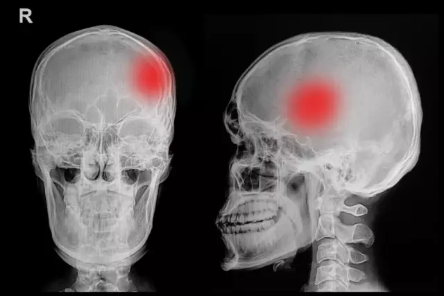
Injuries to the skull can lead to serious disruptions in the functioning of the human body - paralysis, mental disorders, speech and memory impairments. The main traumas of the skull include: fracture of the closed and open vault, fracture of the base of the skull, traumatic brain injury with concussion.
A fracture of the cranial vault manifests itself in the form of a hematoma of the scalp of a person, impaired consciousness, memory loss and respiratory failure. A person who has received this injury should be laid on a flat surface and a bandage should be applied to his head. If the patient is unconscious, it is necessary to put his back on a stretcher in a half-turn position, and put a pillow or roller under one side of the body. In case of breathing disorders, artificial respiration is performed, then the victim is taken to a medical institution for a medical examination.
A skull base fracture can manifest as bleeding from the nose and ears, dizziness and headache, and loss of consciousness. In case of damage to the base of the skull, the victim should free the respiratory tract and oral cavity from cerebrospinal fluid and blood, and in case of respiratory disorders, artificial respiration should be performed.
Concussion occurs in traumatic brain injury. Symptoms are loss of consciousness, dizziness and headache, nausea, vomiting, increased heart rate, pallor of the face, weakness. With a severe brain injury, a person can lose consciousness for several hours. In severe cases, the functioning of the cardiovascular and respiratory systems is disrupted. The victim should immediately be given chest compressions and artificial respiration, and a bandage should be applied to the wound surface, then the patient should be hospitalized.
In the presence of intracranial formations, craniotomy is performed.
Craniotomy is a surgical procedure that creates a hole in the skull bone. The purpose of craniotomy is to reach an injured area that has a hematoma or other malignant formations.
There are several methods of craniotomy - decompression with resection of the temporal bone and opening of the meninges (with dislocation of the bone marrow); osteoplastic with cutting out several soft tissues and bones; resection with removal of a part of the skull bone (for decompression and surgical treatment of brain wounds).
Found a mistake in the text? Select it and press Ctrl + Enter.


